If a veterinarian says a lump on your dog is a hamartoma, the first thing to know is that a hamartoma is benign—it isn’t cancer.
Hamartomas are unusual growths made from tissues that normally belong in that part of the body. Most aren’t dangerous, but there’s an important catch: you can’t tell whether a lump is a hamartoma just by looking at it.
That’s why new or changing lumps should be examined. Depending on the exam, a simple needle sample or biopsy may be recommended to determine exactly what’s going on.
Once a hamartoma is confirmed, treatment is often straightforward. Some can simply be watched, while others are better removed if they’re growing, irritated, bleeding, or bothering the dog.
What exactly is a hamartoma?
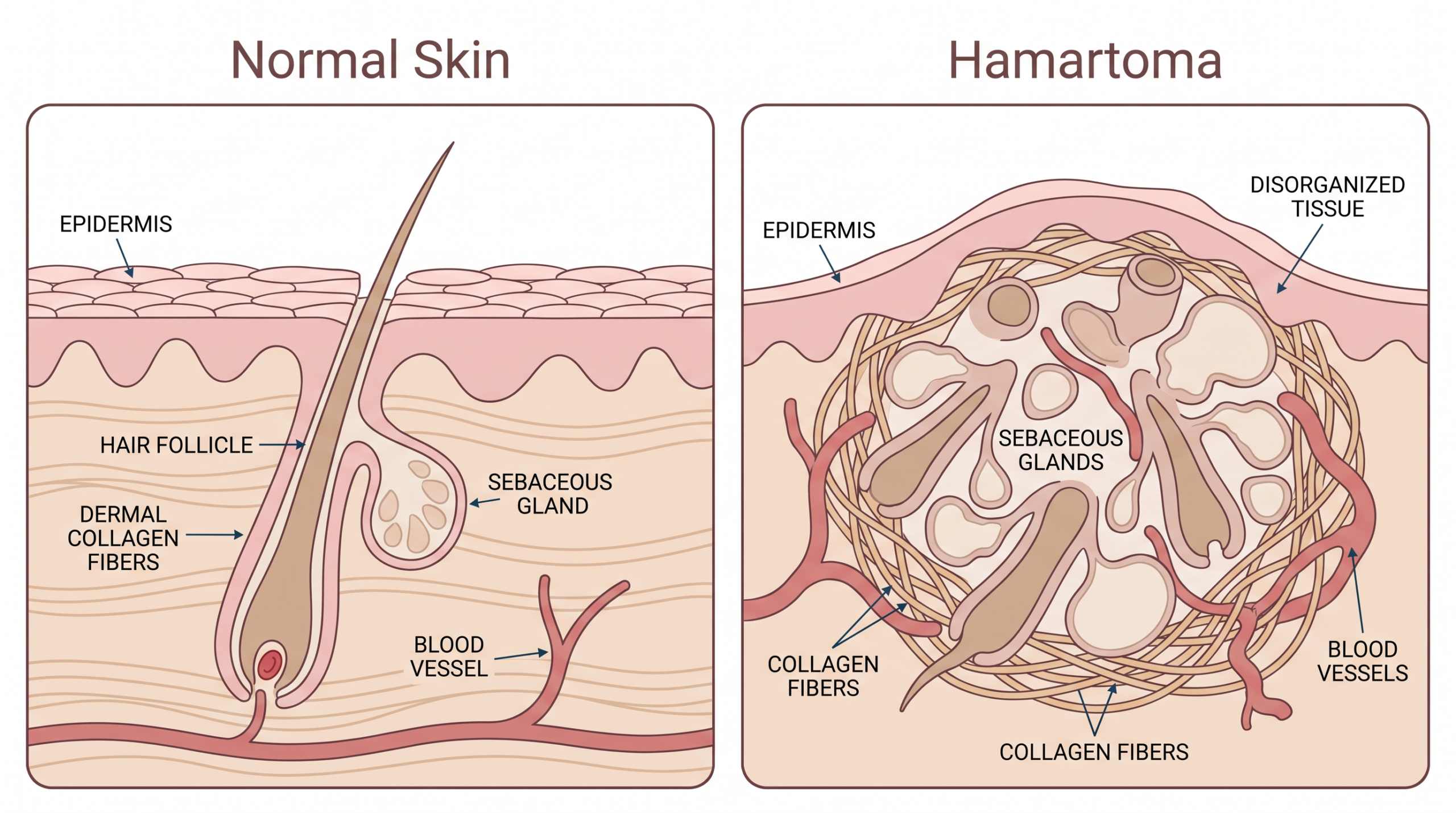
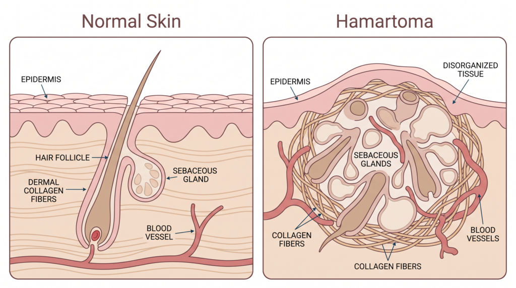
A hamartoma isn’t foreign tissue, and it isn’t a tumor growing out of control. It’s made of the same skin, hair follicles, glands, blood vessels, or connective tissue that already belong in that spot—they’ve just grown together in a disorganized clump instead of in their normal, orderly pattern.
Picture the difference between a well-organized closet and one where everything got shoved in at once. Same items, same materials, just arranged badly. That’s roughly what’s happening under the skin.
Why can’t you just look at it and know?
This is the part every dog owner should understand. Even an experienced veterinarian can’t reliably tell a hamartoma from a more serious growth just by looking at it or feeling it. Size, firmness, and location give useful clues, but they’re not proof. A confirmed hamartoma is not cancer—but an unchecked new lump isn’t a confirmed hamartoma yet, no matter how harmless it looks.
That’s true even for lumps that have been there for months without changing. The only way to know for certain is to look at the cells or tissue itself.
What kinds of hamartomas show up in dogs?
Hamartoma is really an umbrella term. A more specific name is often used depending on which tissues got tangled up, and you might see one of these terms on a pathology report:
| Term on the report | What it means for your dog |
|---|---|
| Fibroadnexal hamartoma | Hair follicles and skin glands bunched together with some extra fibrous tissue. Not cancer. |
| Follicular hamartoma | Extra hair follicles clustered under the skin, usually felt as a firm nodule. |
| Collagenous nevus | A firm bump from a buildup of collagen, a normal connective-tissue protein. |
| Epidermal hamartoma | A darker, slightly rough patch or bump in the top skin layer, most common in young dogs. |
| Vascular hamartoma | Normal blood vessels tangled together instead of running their usual course. |
You don’t need to memorize any of this. If your dog’s report uses one of these terms, your veterinarian can explain exactly what it means for your dog. You might also see the word “nevus” used instead of “hamartoma”—the terms are often used interchangeably.
How a hamartoma is diagnosed
Diagnosis happens step by step rather than by guessing from appearance alone.
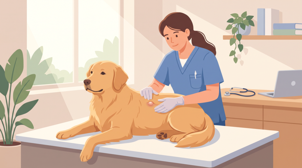
First comes a hands-on exam and a few questions. When was the lump first noticed? Has it changed? Does it bother the dog? This narrows things down, but it’s never enough on its own to name the lump for certain.
Next is often a fine-needle aspirate. This is a quick, well-tolerated test using a small needle to collect a few cells for a closer look under the microscope. It’s great for narrowing possibilities fast, though it doesn’t always give the complete picture.
If a definite answer is needed, a biopsy is the next step. That means removing a small piece of the lump—or the whole thing—and sending it to a lab, where a pathologist examines the actual tissue. This is the only truly reliable way to confirm a hamartoma and rule out anything else it could be.
How a diagnosis comes together
- Step 1 — Exam & history: The lump is examined and its timeline and any changes are discussed.
- Step 2 — Fine-needle aspirate: A quick sample of cells, usually done the same visit.
- Step 3 — Biopsy, if needed: A tissue sample goes to a lab pathologist for a definite answer.
- Step 4 — Treatment plan: Watch it, or remove it — based on the findings and how the lump is behaving.
When should you see your veterinarian?
Any new lump deserves a visit, even if the dog otherwise seems perfectly fine. Getting it looked at early gives the most options and the most peace of mind.
When to see your veterinarian sooner
- The lump is growing or changing shape
- It’s bleeding, oozing, or crusting over
- It looks irritated, red, or infected
- Your dog is licking, chewing, or scratching at it
- It’s in a spot that rubs against a collar, harness, or the ground
None of these automatically mean cancer—but they’re good reasons to get it examined sooner rather than waiting for a routine checkup.
What happens once a hamartoma is confirmed?
There’s no single treatment plan, because hamartomas involve different tissues and show up in different places. Once the diagnosis is confirmed, there are usually two paths: keep an eye on it if it isn’t bothering the dog, or remove it if it’s irritated, infected, growing, or sitting somewhere inconvenient.
Smaller epidermal hamartomas and collagenous nevi typically do well with straightforward surgical removal. Larger or more widespread ones may need a different approach, tailored to the dog’s specific situation.
One important reminder: don’t try to drain, cut, or treat a lump at home. If it is removed, the tissue is usually sent to a lab for testing—that report confirms exactly what it was and whether any follow-up care is needed.
The bottom line
A hamartoma is a benign overgrowth of tissue that already belongs on your dog’s body—it’s just arranged unusually. That’s genuinely good news. But because a hamartoma can look just like something more serious from the outside, any new lump still deserves an exam, and sometimes a biopsy, before you can relax about it.
Frequently asked questions
Are hamartomas in dogs cancer?
No. A hamartoma is a benign growth made of tissue that belongs in that part of the body but grew in a disorganized pattern. Still, get any new lump checked—you can’t tell it’s a hamartoma just by looking at it.
Are hamartomas present at birth?
Often, yes. Hamartomas are developmental, meaning they form as your dog’s body develops rather than appearing later in life. You may just not notice one until it’s grown large enough to see or feel.
What does a hamartoma look like on a dog?
There’s no single look. It can be a small bump, a raised patch, or a firmer nodule depending on the type. Appearance alone can’t confirm the diagnosis—that’s why an exam is always recommended.
Is a collagenous nevus the same thing as a collagenous hamartoma?
Essentially, yes. The terms are often used interchangeably for a firm bump caused by a buildup of collagen in the skin.
How is a hamartoma diagnosed?
Usually starting with a physical exam, often followed by a fine-needle aspirate, and if more certainty is needed, a biopsy sent to a lab for a full look under the microscope.
Does every hamartoma need to be removed?
No. If it isn’t bothering the dog and isn’t in a problem spot, keeping an eye on it may be all that’s needed. Removal is usually reserved for lumps that are growing, irritated, infected, or awkwardly placed.
Should I have my dog’s new lump checked even if it seems harmless?
Yes. A harmless-looking lump can still turn out to need attention, and there’s no way to know for sure without an exam.
Veterinary Sources & References
- Journal of Veterinary Diagnostic Investigation — Cutaneous hamartomas in dogs: case series and classification
- Merck Veterinary Manual — Overview of Tumors of the Skin and Soft Tissues in Animals
- Merck Veterinary Manual — Epidermal and Hair Follicle Tumors in Animals
- VCA Animal Hospitals — Fibrous Tumors: Benign and Hamartomas
- University of Illinois College of Veterinary Medicine — Is That Lump or Bump on Your Pet Cancer?
Comments
One response
[…] a lump where your dog received a vaccine is unsettling, but a small, firm swelling that appears soon after the injection is often a short-lived local reaction. It should begin to […]