Introduction
Mitral valve disease, specifically dog mitral valve disease, is the most common heart disease in dogs, responsible for approximately 80% of heart disease in dogs. This degenerative condition targets the heart’s mitral valve-the critical gateway between the left atrium and left ventricle-causing it to thicken, weaken, and ultimately fail to close properly. While valve disease in dogs can affect any breed, it overwhelmingly impacts small breed dogs and older dogs, with 85% of small-breed dogs developing dog mitral valve disease by age 13.
This guide covers everything dog owners need to know about degenerative mitral valve disease: how it develops, what symptoms to watch for at each stage, how veterinarians diagnose and treat it, and what long-term management looks like. Whether your dog was just diagnosed with a heart murmur or you’re managing congestive heart failure, this resource is designed to help you make informed decisions alongside your veterinary cardiologist.
Understanding dog mitral valve disease is essential for every dog owner. Regular check-ups can help in the early detection of dog mitral valve disease.
Early intervention is crucial in managing dog mitral valve disease to ensure a better quality of life for your pet.
To understand dog mitral valve disease better, consider consulting with a veterinary cardiologist.
Dog mitral valve disease occurs when the mitral valve becomes leaky and thickened through age related degeneration, allowing blood to leak backward from the left ventricle into the left atrium. Left untreated, this mitral regurgitation progressively enlarges the heart and can lead to congestive heart failure.
After reading this guide, you will:
Increased awareness about dog mitral valve disease allows for better care strategies.
Monitoring your dog’s health can help in early detection of dog mitral valve disease.
- Understand how mitral valve disease develops and the stages through which it progresses
- Recognize both subtle early symptoms and emergency warning signs
- Know which diagnostic tests to expect and when to seek veterinary care
- Compare treatment options by disease stage, including medications and mitral valve repair
- Gain practical strategies for home monitoring and long-term quality-of-life management
Understanding Dog Mitral Valve Disease
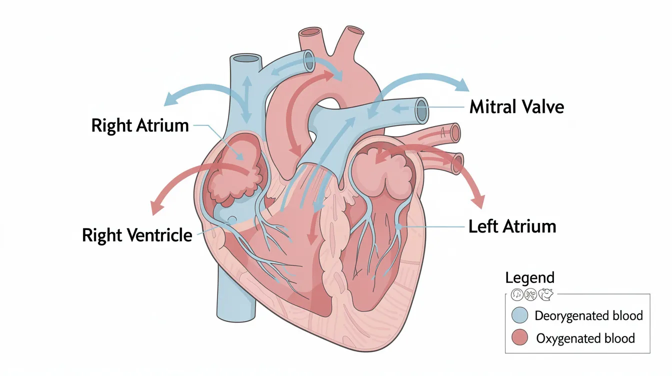
Mitral valve disease is a degenerative condition in dogs where the valve between the heart’s left atrium and left ventricle thickens and leaks. Over time, degenerative changes in the mitral valve cause it to thicken and become floppier, preventing the valve leaflets from sealing tightly. This allows blood leaks backward into the upper left chamber instead of flowing forward to the body. Understanding the anatomy involved, the mechanism of degeneration, and the staging system veterinarians use is essential for every owner navigating this diagnosis.
Canine Heart Anatomy and Function
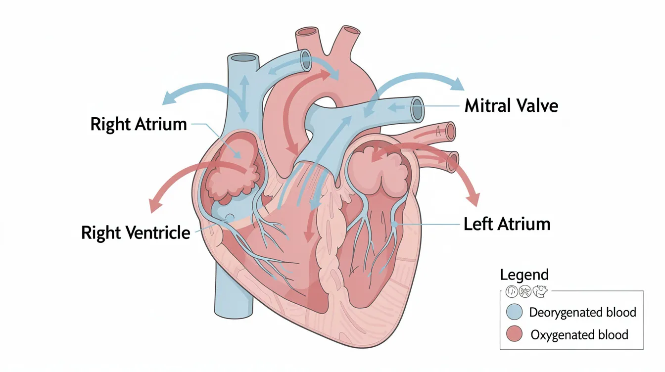
A dog’s heart has four chambers: two upper chambers (the atria) and two lower chambers (the ventricles). The mitral valve sits between the left atrium (the upper left chamber that receives oxygenated blood from the lungs) and the left ventricle (the powerful lower chamber that must pump blood out to the rest of the body).
Many owners find it helpful to learn more about dog mitral valve disease to spot symptoms early.
During normal heart function, the cycle works like this: oxygenated blood fills the left atrium, the mitral valve opens to let blood flow into the left ventricle, and then when the heart contracts, the mitral valve snaps shut so the ventricle can pump blood forward through the aorta to the body. The valve is supported by delicate structures called chordae tendineae (thin cord-like tendons) and papillary muscles, which anchor the valve leaflets and prevent them from flipping backward. When this system works correctly, blood moves efficiently in one direction. When the valve fails, blood can flow backward-and that’s where problems begin.
How Mitral Valve Disease Develops
The primary process behind this heart valve disease is called myxomatous degeneration. The collagen and elastic fibers that give the valve leaflets their strength gradually break down, while abnormal substances (proteoglycans and glycosaminoglycans) accumulate within the valve tissue. The result: leaflets become thickened, stretched, and floppy. This degeneration leads to mitral valve prolapse, where the leaflets bulge backward into the left atrium instead of forming a tight seal.
Make sure to recognize the warning signs of dog mitral valve disease.
Educating yourself about dog mitral valve disease is vital for informed decisions.
Awareness of dog mitral valve disease allows you to be proactive in your pet’s health.
As the disease progresses, the valve’s inability to close properly causes mitral valve regurgitation-blood leaks in the wrong direction with each heartbeat. The heart must then work harder to pump enough blood to the body, causing the heart muscle to enlarge over time. This heart enlargement puts additional strain on the entire cardiovascular system.
Proper knowledge of dog mitral valve disease can lead to timely veterinary visits.
Mitral valve disease has an inherited component in some breeds, with a clear genetic component driving earlier onset. Certain breeds are at higher risk for MVD due to genetics, particularly Cavalier King Charles Spaniels, Dachshunds, Miniature Poodles, Chihuahuas, Maltese, Shih Tzus, and Pomeranians. Cavalier King Charles Spaniels are highly predisposed to mitral valve disease, sometimes showing murmurs as young as four years old-dogs may develop a murmur as early as four years old. Age related changes, body weight, and concurrent health conditions also influence how quickly the disease progresses.
Disease Progression Stages
Mitral valve disease progresses through stages from murmur to congestive heart failure, and veterinarians use the ACVIM (American College of Veterinary Internal Medicine) staging system to classify severity:
- Stage A – Dogs at high risk due to breed predisposition but with no detectable structural heart disease. No murmur, no treatment needed-just awareness.
- Stage B1 – A heart murmur and/or mitral regurgitation is present, but there is no evidence of cardiac enlargement. Dogs in Stages A or B1 require monitoring without medication.
- Stage B2 – Structural changes are now measurable: the left atrium and left ventricle have begun to enlarge. The dog remains asymptomatic, but the heart is remodeling. This is the critical stage where medication may begin.
- Stage C – Clinical signs of congestive heart failure are present or have occurred previously. Fluid in the lungs (pulmonary edema) is common, and active treatment is required.
- Stage D – Advanced, refractory heart failure that no longer responds adequately to standard therapy. Aggressive interventions or palliative care become the focus.
Many dogs live with a murmur for months or even years before any clinical signs appear. Some never advance beyond the earlier stages. Understanding where your dog falls in this system directly shapes treatment decisions-which is why recognizing symptoms early matters so much.
Recognizing Symptoms and Clinical Signs
Do not hesitate to discuss dog mitral valve disease with your veterinarian.
Understanding the implications of dog mitral valve disease is crucial for dog owners.
Because mitral valve disease progresses gradually, symptoms of mitral valve disease often develop so slowly that owners attribute changes to normal aging. Knowing what to look for at each stage can mean the difference between early diagnosis and a crisis visit.
Being informed about dog mitral valve disease leads to better treatment outcomes.
Early Stage Symptoms
Early stages of MVD are often asymptomatic except for a heart murmur. Heart murmurs are the earliest sign of mitral valve disease-a whooshing sound your veterinarian detects during routine auscultation, caused by turbulent blood flow through the leaky mitral valve. At this point, most dogs appear completely normal at home.
Subtle signs that owners sometimes dismiss include slightly decreased stamina on walks, mild reluctance to jump onto furniture, or occasional coughing after exercise. You might notice your dog tiring a bit faster than usual or slowing down in ways you attribute to age. Symptoms may not appear until later stages of the disease, which is why regular veterinary checkups with auscultation are so valuable-veterinarians diagnose MVD through heart murmurs observed during checkups.
Be vigilant for symptoms related to dog mitral valve disease.
Timely recognition of dog mitral valve disease symptoms can save lives.

Advanced Stage Symptoms
As the disease progresses into Stage B2 and Stage C, clinical signs become impossible to ignore. Common clinical signs of MVD include persistent cough and increased breathing rate. The cough is often worse at night or when lying down, caused by the enlarged heart pressing on airways or by fluid in the lungs.
Watch for these advancing symptoms:
- Persistent cough, especially at night or early morning
- Labored or rapid breathing (tachypnea), even at rest
- Exercise intolerance that worsens progressively
- Decreased appetite and gradual weight loss
- Restlessness, particularly difficulty getting comfortable when lying down
- Elevated sleeping respiratory rate-a key indicator that fluid may be accumulating
Secondary mitral regurgitation can also contribute to pulmonary hypertension and, if the tricuspid valve becomes involved, fluid accumulation in the abdomen (ascites). Some dogs develop changes in eating behavior or become increasingly lethargic as heart function declines.
Emergency Warning Signs
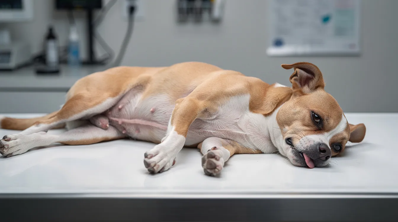
Certain symptoms require immediate veterinary attention-they indicate that heart failure has become life-threatening:
Dog mitral valve disease management includes understanding the clinical signs.
- Severe respiratory distress: open-mouth breathing, audible wheezing or gasping
- Collapse or syncope (fainting episodes)
- Blue or pale gums (cyanosis), indicating inadequate oxygen delivery
- Extreme lethargy or inability to stand
- Sudden, severe cough with possible pink-tinged foam
- Abdominal distention developing rapidly
Pulmonary edema indicates congestive heart failure in dogs and constitutes a medical emergency. In rare cases, the left atrium can rupture under extreme pressure-a catastrophic event. If your dog shows any of these signs, or if you notice hard, labored breathing, seek emergency veterinary care immediately. Recognizing these warning signs and acting quickly can determine whether a dog survives an acute episode.
Recognizing changes related to dog mitral valve disease is important for care.
Diagnosis and Treatment Approaches
Seek veterinary advice if you suspect dog mitral valve disease in your pet.
Accurate diagnosis and proper staging are essential because treatment recommendations differ dramatically between stages. A dog in Stage B1 needs no medication at all, while a dog in Stage C requires multiple daily drugs. Your veterinarian-ideally a veterinary cardiologist for complex cases-will use a combination of physical examination, imaging, and laboratory tests to determine exactly where your dog’s condition stands.
Diagnostic Procedure Steps
Dog mitral valve disease can progress silently; vigilance is essential.
Understanding dog mitral valve disease will allow for better pet care decisions.
When mitral valve disease is suspected (often after detecting a murmur), the diagnostic workup typically follows these steps:
- Physical examination and auscultation – The veterinarian listens for the characteristic holosystolic murmur at the left apex of the chest, grades its intensity (1–6 scale), and checks for abnormal rhythms, jugular vein distension, and overall clinical status. The murmur grade often correlates with severity of mitral insufficiency.
- Chest X rays (thoracic radiographs) – X rays evaluate heart size using the vertebral heart score, identify patterns suggesting fluid in the lungs (pulmonary edema), and establish a baseline for tracking progression. Chest x rays are particularly important for distinguishing Stage B1 from B2 and for confirming congestive heart failure.
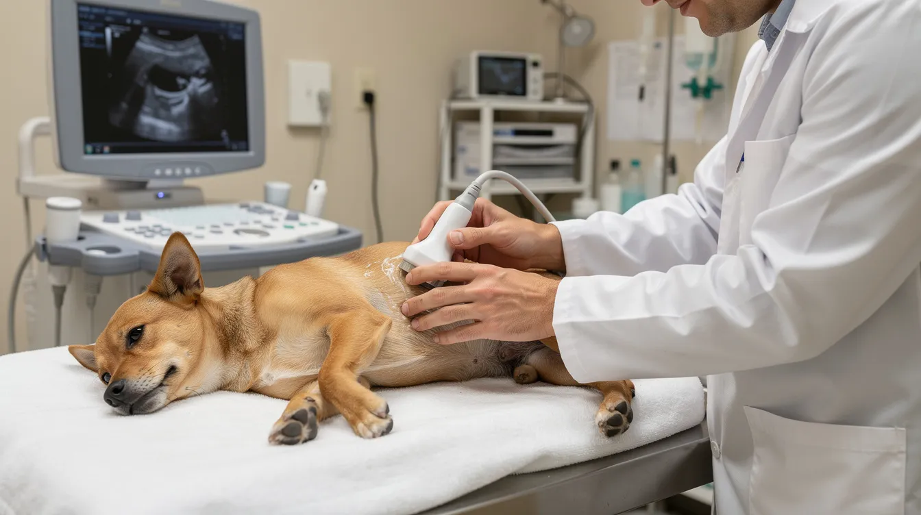
- Echocardiography (cardiac ultrasound) – This is the gold standard for evaluating valve disease in dogs. Regular echocardiograms are essential to monitor the progression of MVD. Using color Doppler imaging, the echocardiogram visualizes valve leaflet thickening, mitral valve prolapse, chordae tendineae integrity, and the direction and volume of regurgitant blood flow. It precisely measures left atrial and left ventricular dimensions-critical data for staging.
- Additional diagnostic tests – ECG (electrocardiogram) detects atrial fibrillation and other abnormal rhythms. Blood work assesses kidney function (essential when prescribing diuretic medications and ACE inhibitors). The biomarker NT-proBNP may be elevated in significant regurgitation or active heart failure. Monitoring blood pressure helps evaluate for high blood pressure or secondary complications.
Breed and age information also factor into the diagnostic picture. Mitral valve disease is more common in small dogs than large breeds, but when larger breeds develop it, they often progress more rapidly and carry a poorer prognosis.
Treatment Options Comparison
Treatment strategies are matched to disease stage. The following table summarizes current evidence-based recommendations:
|
Disease Stage |
Monitoring Frequency |
Medications |
Lifestyle Changes |
|---|---|---|---|
|
Stage B1 (murmur, no enlargement) |
Every 6–12 months; baseline echocardiogram |
No medications indicated; monitoring only |
Maintain lean body weight; moderate exercise; monitor sleeping respiratory rate; avoid high salt treats |
|
Stage B2 (cardiac enlargement, asymptomatic) |
Every 6 months; repeat chest x rays and echocardiograms |
Pimobendan (Vetmedin) to delay onset of CHF; ACE inhibitors (enalapril, benazepril) may be added |
Low-sodium cardiac diet; weight management; adjusted exercise intensity; daily respiratory rate tracking |
|
Stage C (current or prior CHF signs) |
Every few weeks to months depending on stability |
Furosemide (diuretic) to remove excess fluid; pimobendan; ACE inhibitors; spironolactone; oxygen therapy during acute episodes |
Strict low-sodium diet; restful environment; limited strenuous activity; frequent breathing rate checks |
|
Stage D (refractory CHF) |
Very frequent; may require hospitalization |
Increased diuretic doses; combination diuretics (furosemide + torasemide); additional supportive drugs; consider mitral valve repair surgery at specialty centers |
Palliative care focus; comfort-oriented management; tailored nutrition; careful activity limits |
Key medication notes: Medication can manage symptoms of MVD but it cannot be cured. Pimobendan improves heart muscle strength in advanced heart failure and, based on the landmark EPIC trial, significantly delays the onset of CHF when started in Stage B2 dogs. Diuretics like furosemide help remove excess fluid that accumulates due to heart failure. ACE inhibitors like enalapril lower blood pressure in dogs and reduce the workload on the failing heart. All of these medications typically continue lifelong once started, with dose adjustments as the dog’s condition changes.
Surgical option: Heart surgery through mitral valve repair (using artificial chordae and annuloplasty) is increasingly available at specialty centers and has shown improved outcomes in selected patients. A large retrospective study of approximately 1,019 dogs undergoing mitral valve repair (2017–2020) demonstrated that dogs in Stages B2 through D could benefit, though dogs in Stage D had roughly 2.2 times the hazard of early postoperative complications compared to Stage B2 dogs. Factors influencing surgical outcomes included age, body weight, breed (Cavalier King Charles Spaniels carried higher risk than Chihuahuas), and preoperative tricuspid regurgitant velocity.

Common Challenges and Solutions
Living with a dog diagnosed with mitral valve disease presents daily practical challenges. Here are the most common issues owners face and actionable solutions.
Managing Medication Schedules
Dogs in Stage C or D may receive pimobendan twice daily, furosemide two to three times daily, an ACE inhibitor once daily, plus spironolactone-a demanding schedule for any household. Use a weekly pill organizer labeled with your dog’s name and medication times. Set smartphone alarms for each dose. Keep a written medication log to confirm administration, especially when multiple family members share caregiving duties. Watch for side effects including increased thirst and urination (from diuretics), reduced appetite, or signs of dehydration, and report any changes to your veterinarian. Regular blood work monitoring kidney function is essential, as diuretic medications and ACE inhibitors can affect renal health.
Monitoring Disease Progression
Tracking your dog’s sleeping respiratory rate (SRR) at home is one of the most powerful tools available. Count the number of breaths your dog takes in 60 seconds while fully asleep or resting quietly. A consistent rate above 30 breaths per minute may indicate fluid accumulation before obvious symptoms appear.
Interactive Sleeping Respiratory Rate Calculator:
How to use: While your dog is sleeping peacefully, count the number of chest rises in 15 seconds. Multiply that number by 4 to get breaths per minute. Record this daily at the same time. If the rate exceeds 30 breaths/minute on two or more occasions, or if you see a rising trend over several days, contact your veterinarian promptly. Many veterinary clinics also recommend smartphone apps designed for SRR logging that can graph trends over time and alert you to concerning changes.
Also track exercise intolerance patterns, cough frequency and timing, appetite changes, weight fluctuations, and overall energy level. Keeping a simple daily log-even just a few notes on your phone-gives your veterinarian invaluable data for adjusting treatment.
Quality of Life Concerns
Balancing treatment aggressiveness with daily comfort is one of the hardest aspects of managing a leaky heart valve in a senior dog. Medications may extend survival time, but they also introduce pill schedules, potential side effects, and frequent vet visits. Consider these quality-of-life modifications:
- Provide soft, supportive bedding and minimize stairs
- Keep your home at moderate temperature and humidity
- Offer gentle, short walks rather than strenuous exercise
- Maintain a nutritious diet appropriate for cardiac patients
- Obesity can worsen MVD by forcing the heart to work harder-maintaining ideal body weight is critical
Decisions about aggressive interventions like heart surgery versus palliative care should weigh your dog’s current comfort, your family’s capacity, financial considerations, and realistic prognosis. Your veterinarian can help you assess whether your pet’s condition warrants escalation or whether comfort-focused care better serves your dog’s wellbeing.
Conclusion and Next Steps
Early diagnosis improves prognosis and extends lifespan significantly for dogs with mitral valve disease. While this leaky mitral valve condition cannot be cured, proper staging, timely medication, and consistent home monitoring can dramatically improve both survival time and quality of life. Many dogs with mitral regurgitation live comfortably for years with appropriate management-only about 30% of dogs with mitral regurgitation ever develop congestive heart failure.
Your immediate next steps:
- Schedule a veterinary checkup with thorough auscultation, especially if you own a high risk breed or a dog over 7 years old
- Learn to count your dog’s sleeping respiratory rate and begin tracking it daily
- Discuss ACVIM staging with your veterinarian if a murmur has been detected, and ask whether echocardiography is indicated
- Review your dog’s diet for sodium content and discuss cardiac-appropriate nutrition
- Maintain a healthy body weight for your dog-excess weight forces the heart to work harder
Prognosis varies widely based on disease progression and treatment response. Dogs with asymptomatic mitral valve disease live 1 to 5 years, while dogs in congestive heart failure may live 6 to 14 months. For large-breed dogs specifically, median survival time in Stage B1 is approximately 800 days, dropping to around 274 days for Stage B2 and roughly 184 days for Stage C. These are medians, not guarantees-individual outcomes depend on breed, treatment compliance, and how the dog responds to therapy.
Related topics worth exploring include cardiac-specific diets (low sodium with adequate protein and omega-3 fatty acids), exercise modification guidelines for dogs with heart disease, and emerging advances in genetic screening for predisposed breeds.
Frequently Asked Questions
What is the expected lifespan of a dog diagnosed with mitral valve disease? Dogs with asymptomatic mitral valve disease live 1 to 5 years from diagnosis. Dogs in congestive heart failure may live 6 to 14 months with treatment. Survival time depends heavily on the stage at diagnosis, breed, size, and response to treatment. Small breed dogs generally tolerate the disease longer than large breeds when managed properly.
Which breeds are most predisposed to mitral valve disease? Cavalier King Charles Spaniels are the most predisposed breed, often developing murmurs earlier than other dogs. Other high risk breeds include Dachshunds, Miniature Poodles, Chihuahuas, Maltese, Shih Tzus, Miniature Schnauzers, and Pomeranians. Mitral valve disease is more common in small dog breeds, and 85% of small-breed dogs will have mitral valve disease by age 13. Males are slightly more commonly affected than females.
Is there a cure for mitral valve disease in dogs? No. Mitral valve disease is a degenerative condition that cannot be reversed. Medication can manage symptoms of MVD but it cannot be cured. Mitral valve repair surgery can significantly improve heart function in selected patients, but it is expensive, available only at specialty centers, and carries surgical risks. All treatments are aimed at slowing progression, managing symptoms, and maintaining quality of life.
How much does treatment cost? Costs vary by location and disease stage. Diagnostic echocardiograms and chest x rays may range from several hundred to over a thousand dollars. Monthly medication costs (pimobendan, diuretics, ACE inhibitors) depend on dog size and number of drugs required. Surgical mitral valve repair at specialty hospitals is substantially more expensive. Ongoing monitoring adds to lifetime costs.
When should medication be started? Dogs in Stages A or B1 require monitoring without medication-no drugs have been proven beneficial at these earlier stages. Pimobendan is recommended starting at Stage B2 (when cardiac enlargement is documented) based on the EPIC trial, which showed it significantly delays the onset of congestive heart failure. Diuretics and ACE inhibitors are typically added when clinical signs of heart failure emerge at Stage C.
Can diet and lifestyle slow disease progression? Yes. Low salt diets may help prevent fluid retention in dogs with cardiac disease. Avoiding high salt treats, maintaining ideal body weight, and providing regular moderate exercise (rather than strenuous activity) all support heart function. Omega-3 fatty acid supplementation and adequate protein intake are also recommended as part of a cardiac management plan.
How do I monitor my dog at home? Counting sleeping respiratory rate is the single most valuable home monitoring tool. A normal sleeping rate is typically under 30 breaths per minute. Track this daily and report any sustained increases to your veterinarian. Also watch for changes in cough frequency, exercise intolerance, appetite, and overall energy. Some owners use wearable activity trackers or smartphone apps to log trends over time.
Dog mitral valve disease may not show symptoms until advanced; regular checks help.
What complications should I watch for? Congestive heart failure can develop over several years, but owners should also watch for atrial fibrillation (irregular heart rhythm), worsening kidney function (especially on diuretics and ACE inhibitors), and rare but serious events like chordae tendineae rupture or left atrial tear. Any sudden change in your pet’s condition-especially in breathing, energy level, or gum color-warrants immediate veterinary evaluation.
Should dogs with mitral valve disease be bred? In high risk breeds, breeding dogs should be screened with auscultation and echocardiography. If a murmur or echocardiographic evidence of mitral regurgitation appears before age 6–8 years, most guidelines recommend retiring that dog from breeding programs. Because mitral valve disease has an inherited component in some breeds, responsible breeding decisions can reduce disease prevalence in future generations.
What is the difference between mitral valve disease and mitral valve stenosis? Mitral valve disease (specifically degenerative or myxomatous mitral valve disease) involves valve thickening and leaking-the valve fails to close properly and blood flows backward. Mitral valve stenosis, which is rare in dogs, involves narrowing of the valve opening that restricts forward blood flow. The vast majority of canine mitral valve conditions are degenerative rather than stenotic.
Additional Resources
Reviewed by Dr. Roger Hart, DVM
References:
- Merck Veterinary Manual. “Myxomatous Atrioventricular Valve Degeneration in Dogs and Cats.” Reviewed January 2023, modified May 2025. Comprehensive clinical reference covering pathophysiology, diagnosis, and treatment principles for degenerative valve disease.
- Cornell University College of Veterinary Medicine. “Leaky Valve Disease of Older Dogs.” Department of Cardiology. Clinical overview of degenerative atrioventricular valve disease including clinical signs, diagnostics, treatment approaches, and prognosis for affected dogs.
Mitral valve disease is manageable with the right combination of monitoring, medication, and regular veterinary follow-up – early detection and consistent care give dogs the best long-term outcome.