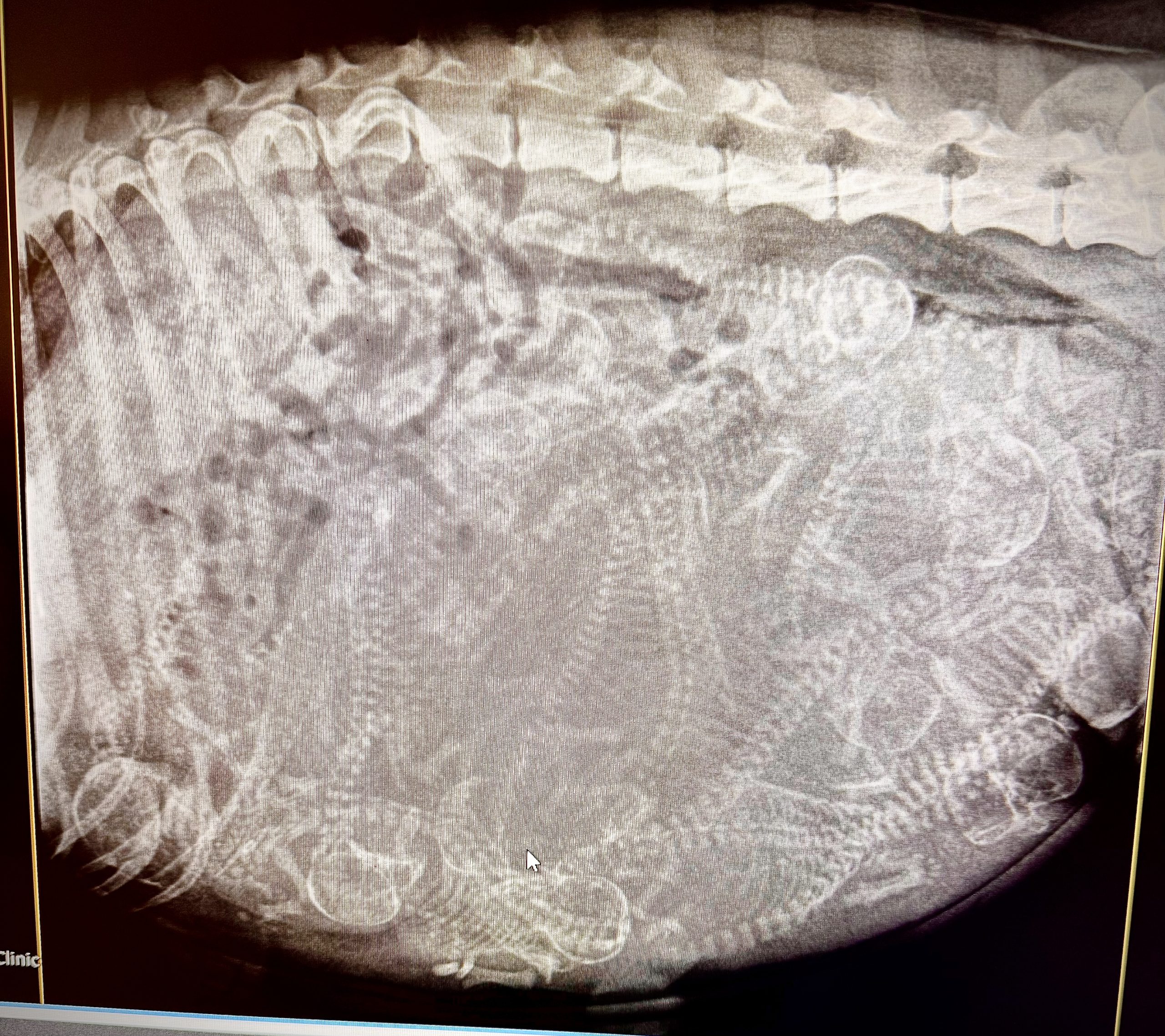
Introduction
X-rays are the most reliable way to count puppies and check delivery readiness in a pregnant dog, but timing matters. Puppy skeletons don’t show up clearly on radiographs until around day 45 of pregnancy, and the most accurate count comes from images taken after day 55. Ultrasound is better for confirming pregnancy early. A single diagnostic x-ray taken at the right stage carries only a tiny radiation dose and is considered safe for the mother and her litter.
This article covers what pet owners need to know about pregnant dog x-rays: the right timing for each goal, radiation safety, what happens during the procedure, and how the results support a safer delivery. Ultrasound plays an important role early in pregnancy too, and the sections below explain how the two imaging methods work together across gestation.
Whether you’re a first-time breeder or a concerned pet owner whose dog is expecting, this guide is written for you. Understanding when and why to schedule a pregnancy x-ray helps you make informed decisions about both the mother and her litter, in partnership with your veterinarian.
What you’ll learn from this article:
- Which imaging method and timing fits your goal, from confirming pregnancy to planning for delivery
- Why the day 45–55 window matters for fetal skeletal visibility
- How radiation exposure from pregnancy x-rays compares to safe thresholds
- Step-by-step preparation for the procedure
- How accurate puppy counts reduce whelping complications
- The specific signs that mean whelping has become an emergency
Which Imaging Timing Fits Your Goal?
Not every pregnant dog needs an x-ray on the same day. The right test and the right timing depend on the goal: confirming pregnancy, checking on the puppies, counting the litter, or planning for delivery. Use this table as a quick reference before the details that follow.
| Goal | Best Method | Recommended Timing | Why |
|---|---|---|---|
| Confirm pregnancy | Ultrasound | Day 25–30 after breeding | Detects gestational sacs long before puppy skeletons appear on an x-ray |
| Check fetal heartbeats | Ultrasound | Day 25 onward | Shows viability directly, which an x-ray cannot do |
| See the first skeletal detail | X-ray | Day 45 onward | Bones absorb enough radiation to appear on film starting around day 45 |
| Get an accurate puppy count | X-ray | Day 55 or later | Skulls and spines are fully mineralized and countable |
| Check for a size mismatch before delivery | X-ray | Final week of pregnancy | Compares puppy skull size to the mother’s pelvis |
| Confirm whelping is finished | X-ray | As soon as a retained puppy is suspected | Compares the pre-whelping count to the puppies already delivered |
Understanding Pregnancy Imaging in Dogs
Radiographic imaging during canine pregnancy uses a low dose of ionizing radiation to create detailed two-dimensional images of the abdomen and uterus. X-rays pass through soft tissue but are absorbed by mineralized bone, which is why they become invaluable once the developing puppies’ skeletons have calcified enough to appear on film. For veterinarians, this imaging method serves a specific purpose: determining how many puppies are present, assessing fetal size and position, and identifying potential complications before whelping begins.
For expectant dog owners and breeders, understanding the role of pregnancy radiographs removes much of the uncertainty surrounding birth. Knowing the litter size ahead of time helps prepare supplies, arrange veterinary support, and recognize if a puppy has been retained during delivery. A retained puppy can become life-threatening without prompt intervention, and it is one of the reasons newborn puppies are lost around whelping.
X-ray vs. Ultrasound: Quick Comparison
Two primary imaging methods are used during a dog’s pregnancy, and each serves a distinct purpose at different stages of gestation.
| Method | Best Used | Strengths | Limitations |
|---|---|---|---|
| Ultrasound | Days 25–35 (early pregnancy) | No radiation, confirms pregnancy and fetal heartbeats, and estimates gestational age | Less accurate for counting puppies, especially in large litters |
| X-ray | Day 45+ (skeletal mineralization), most accurate after day 55 | Most reliable puppy count, shows skull size vs. maternal pelvis, and supports whelping and C-section planning | Cannot confirm pregnancy in the first three weeks. Overlapping skeletons can complicate counts in very large litters |
Ultrasound (ultrasonography) uses sound waves rather than radiation, making it the preferred method to confirm pregnancy early, typically between days 25 and 35 after breeding. It can detect fetal heartbeats, assess viability, and help calculate gestational age based on crown-rump length measurements. Ultrasound is less accurate for counting puppies than x-rays, particularly when a large number of fetuses are present and overlap within the uterus.
X-rays (radiography) become the superior method later in pregnancy, after fetal bones have mineralized sufficiently to be visible. X-rays can determine the number of puppies in a litter with far greater precision than ultrasound, and they provide a clear view of puppies’ bone structures, including skull size relative to the mother’s pelvis. This information is critical for assessing whether a natural birth is feasible or a cesarean section is needed.
In most cases, veterinarians recommend ultrasound first for early pregnancy diagnostics, then x-rays later for puppy counting and pre-whelping planning.
Radiation Safety Fundamentals
One of the most common concerns among pet owners is whether x-rays are safe for a pregnant dog. The answer is reassuring. Diagnostic benefits clearly outweigh the risk when the study is done at the right stage, a conclusion supported by both veterinary and human medicine research.
A single x-ray delivers approximately 30 to 50 mrem of radiation, a dose far below any known harm threshold. Veterinary radiology safety reviews put 0.05 Gy (50 mGy) as the exposure level below which no increased pregnancy risk is expected, and a properly performed diagnostic radiograph stays well under that benchmark.
The stage of pregnancy also matters. Early gestation, when organogenesis and critical development occur in roughly the first three weeks, is the period of greatest vulnerability to radiation. By the time x-rays are typically performed, after day 45, the puppies have progressed beyond this sensitive stage, and their tissues are substantially more resistant to radiation effects.
With this safety foundation established, the next question becomes: exactly when should a pregnancy x-ray be scheduled?
Optimal Timing for Pregnant Dog X-rays
Timing depends on the goal. The sections below walk through each stage of pregnancy and what an x-ray, or an ultrasound, can and cannot show at that point.
Confirming Pregnancy: Ultrasound Comes First
Ultrasound remains the better tool for early pregnancy questions. It can detect gestational sacs by around day 25 to day 30 after breeding and can confirm fetal heartbeats from about day 25 onward. An x-ray taken this early shows nothing useful, because fetal bones have not yet mineralized enough to appear on film. If the goal is simply confirming pregnancy or checking that the puppies are alive, an ultrasound is the right test, not an x-ray.
Day 45: The Earliest Useful X-ray
Fetal skeletal mineralization, the process by which puppy bones accumulate minerals and become dense enough to appear on radiographs, begins around day 42 of gestation. The bones are not reliably visible on standard x-rays until approximately day 45 to 48. Before this point, the developing puppies’ skeletons are not dense enough to absorb x-rays differently from the surrounding soft tissue, so radiographs taken before day 45 usually show nothing diagnostic, even in a confirmed pregnancy.
X-rays taken around day 45 mark the earliest point for skeletal detection, though images from this window can still show incomplete mineralization. The Merck Veterinary Manual notes that developing fetuses can be detected on x-rays after about day 45, with clearer detail as mineralization progresses through day 55.
If breeding dates are uncertain, as they often are when ovulation timing was not tracked precisely, a veterinarian can use palpation findings, hormone assays, or earlier ultrasound measurements to estimate gestational age before scheduling the x-ray.
Day 55 and Later: The Best Window for an Accurate Count
X-rays are most accurate in the final week of pregnancy. After day 55, skeletal detail is well defined, which allows the most accurate puppy count possible. The Merck Veterinary Manual states that x-rays taken more than 55 days into pregnancy are the best way to determine litter size.
At this stage, radiographs do more than answer how many puppies to expect:
- Fetal size assessment: X-rays allow measurement of puppy size relative to the mother’s pelvis, which matters most in brachycephalic breeds and dogs with narrow pelvic canals
- Positional evaluation: Identifying whether puppies are in normal head-first or breech positions helps anticipate delivery complications
- C-section planning: A puppy-count x-ray helps identify the need for a C-section due to large puppies, an oversized litter, or anatomical concerns in the mother
Knowing the exact number of fetuses present is the most practical benefit. During whelping, this number shows whether all puppies have been delivered or if one remains in the uterus, a situation that can lead to dystocia (a difficult or obstructed birth) or uterine infection. Any doubt about whether whelping is complete calls for a prompt check from a veterinarian.
Assessing Delivery Risk and Planning a C-Section
A late-pregnancy x-ray does more than count puppies. It also lets a veterinarian compare each puppy’s skull size to the width of the mother’s pelvic canal. A size mismatch raises the odds of a difficult, obstructed birth.
This assessment carries extra weight for breeds with large heads relative to body size, including Bulldogs, French Bulldogs, and Boston Terriers. When a mismatch is found before labor starts, a scheduled cesarean section can be planned in advance, rather than attempted as an emergency once labor has already stalled.
| Call for veterinary help now if: |
|---|
| Strong contractions continue for more than 20 to 30 minutes without a puppy being delivered |
| More than 2 to 4 hours pass between puppies with no active contractions |
| Weak or infrequent contractions continue for more than 1 to 2 hours with no progress |
| A puppy is visibly stuck in the birth canal |
| Green, dark, or foul-smelling vaginal discharge appears before all puppies are delivered |
| Heavy bleeding, a fever above 103°F, or signs of severe pain or exhaustion appear |
| The pre-whelping x-ray count has not been reached and no further puppies are being delivered |
Emergency Situations
There are circumstances when x-rays are needed regardless of optimal timing. If a pregnant dog experiences trauma, shows signs of fetal distress, or develops symptoms suggesting a complication such as pyometra, urgent imaging can be warranted even before day 45.
In emergency scenarios, the diagnostic benefit of assessing the abdomen for pelvic fractures, foreign bodies, or fetal abnormalities outweighs the minimal radiation risk. Veterinary teams apply strict safety protocols to minimize exposure while getting the information needed.
Key timing takeaways:
- Day 42–44: Mineralization begins but usually is not visible on radiographs
- Day 45–48: Earliest reliable skeletal detection, and the earliest point an x-ray should be performed
- Day 55–60: Best window for an accurate puppy count and pre-whelping assessment
- Final week: Best window to compare fetal size against the mother’s pelvis and plan for a possible C-section
- Emergency: Any time clinical urgency outweighs timing considerations
The X-ray Procedure and Safety Protocols
With a clear understanding of when to schedule the x-ray, here’s what to expect during the actual procedure and how veterinary teams ensure safety for both the mother and her developing puppies.
Preparation Steps
A veterinarian can provide specific instructions before the appointment, but general preparation follows a consistent pattern:
- Pre-appointment fasting: Many clinics recommend withholding food for several hours before the x-ray. An empty stomach reduces gas and food-related artifacts in the abdomen, producing clearer images and reducing the chance of needing repeat exposures. A veterinarian will specify the exact fasting window.
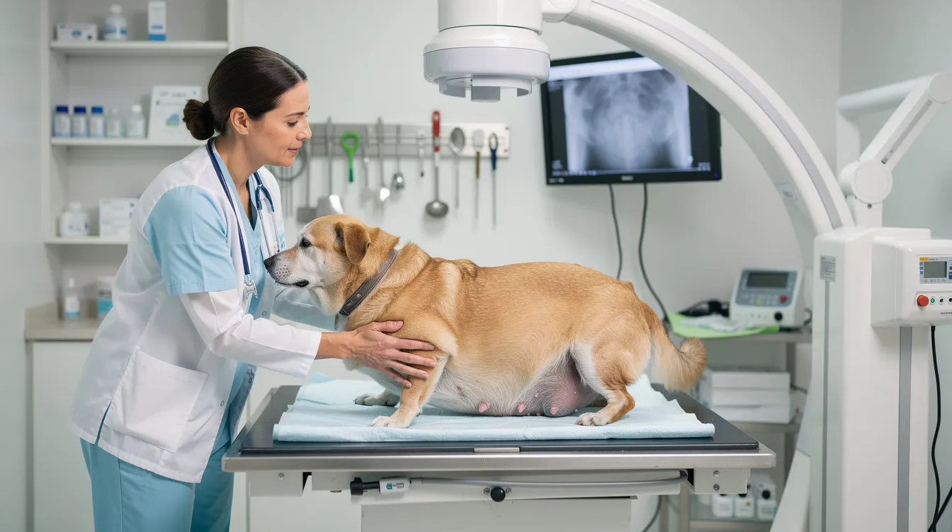
- Comfort measures for the pregnant dog: A calm environment significantly improves image quality. Soft padding on the x-ray table, gentle handling, and familiar blankets or toys help reduce stress. Anxious dogs are more likely to move during imaging, which degrades the radiograph and can require additional exposures.
- Positioning considerations for optimal images: The dog is typically positioned in two views, lateral (lying on her side) and ventrodorsal (lying on her back). Both views are essential for an accurate count because fetuses that overlap in one orientation can be distinguishable in the other. Foam wedges and sandbags help maintain proper alignment without requiring hands-on restraint from staff.
- Sedation assessment and safety protocols: Most pregnant dogs tolerate the procedure without sedation. If a dog is extremely anxious or cannot remain still, light sedation can be considered. A veterinarian will select agents with established safety profiles for pregnant patients, using the minimum effective dose and monitoring closely throughout.
Radiation Safety Comparison
Understanding how the radiation from a pregnancy x-ray compares to other exposure sources helps put safety into perspective:
| Exposure Source | Approximate Dose | Risk Level |
|---|---|---|
| Single pregnancy x-ray | 30–50 mrem (0.3–0.5 mSv) | Negligible, well below harm threshold |
| Annual background radiation (human reference) | ~310 mrem (3.1 mSv) | Normal environmental exposure |
| Harmful threshold for pregnancy | 5,000 mrem (50 mSv / 0.05 Gy) | Level below which no increased risk expected |
As this comparison shows, the radiation involved in a diagnostic abdominal radiograph is extremely low, delivering roughly 1/100th of the exposure level at which any pregnancy risk has been documented. Modern digital radiography systems require lower doses than older film-based equipment, adding another margin of safety.
Veterinary staff protect themselves with lead aprons and thyroid shields during the procedure, and pregnant veterinary personnel are typically excused from direct involvement in radiographic imaging to reduce unnecessary exposure, even though appropriately used x-rays are considered safe for patients.
Common Challenges and Solutions
Even with careful planning, several practical concerns frequently arise when pet owners consider pregnancy x-rays for their dogs.
Dog Anxiety During the Procedure
Some dogs, particularly those unfamiliar with clinical environments, become stressed during positioning and imaging. Movement during exposure produces blurred radiographs, potentially requiring repeat images and additional radiation exposure.
Solution: Low-stress handling techniques make a significant difference. Allow the dog a few minutes to acclimate to the room. Use treats and calm verbal reassurance. Request that the same handler stay with the dog throughout. If anxiety is severe despite these measures, discuss pregnancy-safe sedation options with a veterinarian.
Unclear Litter Size and Puppy Count Results
Even with optimal timing, overlapping skeletons can make it difficult to achieve a definitive count, especially in dogs carrying a large number of puppies. A lateral view can show seven skulls while a ventrodorsal view suggests eight, leaving uncertainty.
Solution: Two orthogonal views (lateral and ventrodorsal) are standard practice precisely because they reduce ambiguity. If the count remains unclear, an experienced veterinary radiologist can review the images. In some cases, combining late-term x-ray findings with earlier ultrasound estimates provides the most reliable assessment. A veterinarian can note a range instead, for example seven to eight puppies, and advise monitoring during delivery accordingly.
Cost Questions
Pregnancy x-rays represent an additional expense during what can already be a costly breeding process. Costs vary by location, clinic, and the number of views required. Pregnancy radiographs commonly range from about $150 to $300, and an earlier ultrasound assessment typically costs $100 to $200. Exact pricing depends entirely on the local veterinary practice.
Solution: Ask for a cost estimate before the appointment. Many practices offer whelping preparation packages that bundle imaging with prenatal consultations. Ask whether pet insurance covers reproductive diagnostics, since some policies include breeding-related care while others exclude it. Consider the practical value: knowing puppy count helps prevent complications during delivery, potentially avoiding far more expensive emergency interventions.
Conclusion and Next Steps
Pregnant dog x-rays are a safe, valuable diagnostic tool when performed at the right stage of gestation, and at the stage that matches the owner’s specific goal. The key points to remember: x-rays should be performed after day 45 of pregnancy, with the most accurate results achieved after day 55. Radiation exposure is negligible compared to established safety thresholds, and the information gained, including an accurate puppy count, fetal size assessment, and positioning evaluation, directly supports safer whelping outcomes for both the mother and her puppies.
Your next steps:
- Confirm gestational age with a veterinarian using breeding dates, palpation, or ultrasound findings
- Match the timing to the goal using the table above, whether that’s early skeletal detection, an accurate puppy count, or delivery-risk assessment
- Prepare questions about sedation needs, fasting instructions, and what the images will reveal
- Watch for pregnancy signs in the final week: nesting behavior, a temperature drop, and reduced appetite often signal approaching labor
- Learn the signs of a whelping emergency, covered above, so help can be requested without delay
For comprehensive whelping preparation, set up a birth plan with a veterinarian that accounts for the x-ray findings. If the radiographs suggest potential complications, such as oversized puppies, abnormal positioning, or a mismatch between fetal skull size and maternal pelvis, a veterinarian can outline contingency plans, including surgical delivery if needed.
Combining both imaging methods across pregnancy, ultrasound early and x-rays late, gives the most complete picture of fetal health and delivery readiness.
Frequently Asked Questions
Can x-rays harm my pregnant dog or her puppies?
When performed after skeletal mineralization (approximately day 45) with proper safety protocols, x-rays are considered safe for most pregnant dogs. Diagnostic x-rays deliver doses far below the 0.05 Gy threshold at which any increased risk has been documented, and the long-term cancer risk from a properly performed diagnostic study is considered negligible. In nearly every case, the diagnostic benefit substantially outweighs the extremely low radiation risk. A veterinarian is still best positioned to weigh this for a specific dog’s situation.
How accurate are x-rays for counting puppies?
X-rays taken after day 55 provide the most accurate puppy count available through any imaging method. Accuracy can vary with litter size. In very large litters, overlapping skeletons can cause counts to be off by one or two. Taking two orthogonal views (lateral and ventrodorsal) improves precision. Ultrasound is less effective than x-rays for counting puppies, particularly when many fetuses are present.
What’s the difference between x-rays and ultrasound for pregnant dogs?
Ultrasound uses sound waves (no radiation) and excels at early pregnancy confirmation (days 25–35), detecting heartbeats, and assessing fetal viability. X-rays use ionizing radiation and are performed later (day 45+) to count puppies, evaluate bone structures, and assess fetal size relative to the birth canal. The two methods are complementary: ultrasound for early detection and viability, x-rays for late-term counting and delivery planning.
Should the x-ray be scheduled for puppy count, or is it fine to just wait and see how many are born?
Waiting to see how many puppies arrive naturally makes it hard to know when whelping is actually finished. A retained puppy can be missed without a known target number, and that mistake can become an emergency. A pre-whelping x-ray after day 55 gives a specific number to watch for, which is the main reason most veterinarians recommend scheduling one instead of guessing.
Do I need to do anything special before my dog’s pregnancy x-ray?
Know the breeding dates if possible, since this helps determine optimal timing. A veterinarian can recommend light fasting before the appointment to reduce abdominal artifacts. Keep the dog calm and comfortable, and bring a familiar blanket if it helps. A veterinarian will handle positioning and safety protocols, but questions about sedation plans and what to expect during the procedure can be asked in advance.
When is the earliest I can get an x-ray to count puppies?
The earliest reliable detection of fetal skeletons on radiographs occurs around day 45 of gestation, when bone mineralization becomes visible. For the most accurate count, waiting until after day 55 is strongly recommended. X-rays can determine the number of puppies in a litter most reliably during this later window, when skeletal detail is fully developed.
How much do pregnancy x-rays for dogs typically cost?
Costs vary by geographic location, clinic type, and the number of views needed. Pregnancy radiographs commonly range from about $150 to $300, with earlier ultrasound assessments running roughly $100 to $200. A veterinarian can give specific pricing directly, and many practices offer bundled whelping preparation packages that include imaging along with prenatal consultations.
Should I get x-rays if my dog seems healthy during pregnancy?
Even in a healthy, uncomplicated pregnancy, x-rays provide valuable information. They help confirm the number of puppies in a litter, which is essential for knowing whether all puppies have been delivered during whelping. They also reveal fetal size and position, helping identify potential delivery complications before they become emergencies. For breeders or owners of breeds prone to dystocia, pre-whelping radiographs are considered standard practice.
Can small breed dogs safely get x-rays during pregnancy?
Yes, x-rays are safe for pregnant dogs of all sizes when protocols are followed. Small breed dogs can actually benefit more from pre-whelping radiographs because their smaller pelvic dimensions increase the risk of size mismatch between puppies and the birth canal. Positioning can require extra care in small patients, and overlapping skeletons can be harder to differentiate, but the radiation safety profile remains the same regardless of breed size.
Additional Resources
For further guidance on canine pregnancy management and whelping preparation, these related guides can help:
- What Kills Puppies: Critical Threats Every Owner Must Know: understanding whelping and neonatal risks that make pre-whelping imaging valuable
- Canine Pyometra: How to Know if This is Critical?: recognizing a separate uterine emergency that can sometimes cause confusion around pregnancy symptoms
- Puppies Vaccines Cheap: What You Need to Know: planning ahead for the new litter’s first veterinary visits
Keep a veterinarian’s contact information readily accessible as the due date approaches, and set up a whelping plan that incorporates the x-ray findings. If labor stalls, a puppy seems retained, or the dog shows signs of distress, contact a veterinarian or the nearest emergency veterinary hospital right away.
Veterinary Sources & References
- Merck Veterinary Manual, Pregnancy Determination in Bitches and Queens
- Merck Veterinary Manual, Dystocia in Small Animals
- Schrank M, Pereira M, Mollo A. Diagnostic imaging in the pregnant bitch: risks, advantages, and disadvantages. Frontiers in Veterinary Science, 2025. Full text via PMC
- American College of Veterinary Radiology, ACVR’s Radiation Safety Statement
- Cornell University College of Veterinary Medicine, Riney Canine Health Center, Dystocia in Dogs: Understanding Difficult Births