Introduction
Glaucoma in dogs is a serious eye disease characterized by increased intraocular pressure (IOP) that damages the optic nerve and retina, often leading to irreversible blindness if left untreated. This condition represents a true veterinary emergency – permanent vision loss can begin within hours of acute pressure elevation, making early diagnosis and immediate treatment essential for any chance of preserving your dog’s sight.
This comprehensive guide covers everything pet owners need to know about canine glaucoma: what causes the pressure inside the eye to rise, which breeds are genetically predisposed, how to recognize both subtle signs and advanced symptoms, the full range of medical and surgical treatment options available today, and what realistic outcomes look like. Whether your dog has just been diagnosed or you’re researching because your breed is at higher risk, this guide will help you make informed decisions alongside your veterinarian.
Glaucoma in dogs is a condition where inadequate drainage of fluid called aqueous humor causes dangerously increased pressure within the eye, requiring urgent veterinary attention to prevent permanent vision loss.
By reading this guide, you will:
- Understand the difference between primary and secondary glaucoma and why it matters for your dog’s treatment plan
- Learn to recognize the early warning signs of glaucoma before irreversible damage occurs
- Compare medical treatment and surgical options with current success rate data
- Know what to expect for long-term prognosis and quality of life management
Understanding Glaucoma in Dogs
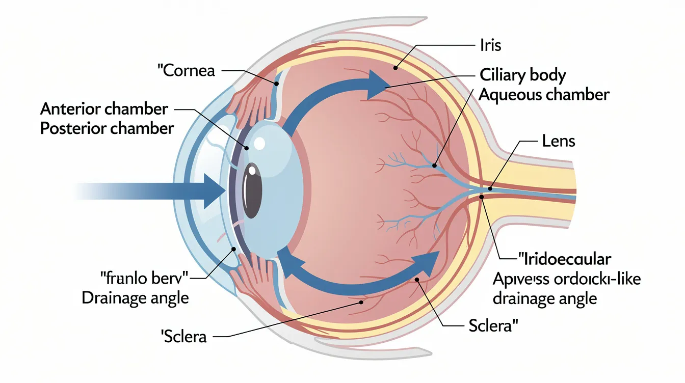
Glaucoma is defined as a pathologic elevation of intraocular pressure IOP that damages the structures responsible for vision – primarily the optic nerve and retina. In a healthy dog eye, the ciliary body continuously produces a clear fluid called aqueous humor. This fluid flows from the posterior chamber, through the pupil, into the anterior chamber, where it nourishes the lens and cornea before draining out through the iridocorneal angle (also called the drainage angle). In dogs, approximately 85% of aqueous humor exits through this conventional pathway, with the remaining 15% draining through the uveoscleral route.
When this drainage system malfunctions – whether due to an anatomical abnormality, inflammation, or physical blockage – excess fluid accumulates and pressure builds. This increased IOP compresses delicate retinal ganglion cells and optic nerve fibers, progressively destroying them. According to the University of Missouri College of Veterinary Medicine, early treatment is critical because permanent vision loss can begin within hours of dangerous pressure elevation.
How Intraocular Pressure Affects Your Dog’s Vision
Normal intraocular pressure in dogs typically ranges between 15 and 25 mmHg, with readings up to approximately 28 mmHg sometimes falling within normal limits depending on the measurement device and breed. Values consistently above this range are considered abnormal, and IOP above 40–50 mmHg requires emergency treatment.
When pressure rises beyond normal thresholds, the mechanical force compresses blood vessels supplying the retina and physically damages the optic nerve head – a process called “cupping.” Retinal ganglion cells are particularly sensitive to this pressure; once destroyed, they do not regenerate. Increased intraocular pressure can lead to blindness, and chronic elevation causes the globe itself to stretch and enlarge, a condition called buphthalmos. This structural damage is permanent and cumulative, which is why even brief periods of uncontrolled pressure can have devastating consequences for your dog’s vision.
Breeds at Higher Risk
Primary glaucoma is often hereditary and breed-related. Certain breeds carry genetic predispositions that affect the anatomy of their drainage angle, making them significantly more likely to develop glaucoma. High-risk breeds include Cocker Spaniels, Basset Hounds, Beagles, Boston Terriers, French Bulldogs, Chow Chows, Shar Peis, and many terrier breeds. In a recent cyclocryotherapy study, Shih Tzus were disproportionately represented among affected dogs, and female dogs showed a higher incidence ratio of approximately 1.67:1 compared to males in primary glaucoma cases.
These breeds often have inherited abnormalities of the iridocorneal angle – narrow, collapsed, or malformed pectinate ligaments – that progressively restrict aqueous humor outflow. Zonular weakness, which predisposes to lens luxation, and brachycephalic eye conformation add further risk. Understanding your dog’s breed-related risk is the first step toward appropriate screening and early intervention, which connects directly to whether your dog may face primary or secondary glaucoma.
Types of Glaucoma in Dogs
Whether rooted in inherited anatomy or triggered by another eye condition, glaucoma is classified as primary or secondary glaucoma based on its underlying cause. This distinction matters because it directly influences treatment strategy, expected outcomes, and whether the other eye is also at risk.
Primary Glaucoma
Primary glaucoma is an inherited condition where congenital or early-onset abnormalities of the drainage angle block aqueous humor outflow independent of any other eye disease. Primary angle-closure glaucoma (PACG) is the most common form in dogs, typically presenting in middle-aged to older purebred dogs. The condition tends to affect one eye first, but because the anatomical abnormality is bilateral, the second eye almost always develops glaucoma eventually – often within months to a couple of years.
Recent research has revealed that even subclinical inflammation may play a role in accelerating progression in primary glaucoma cases, suggesting that the disease involves more than simple mechanical obstruction. Genetic studies continue to identify risk alleles in predisposed breeds such as Cocker Spaniels and Basset Hounds, with the goal of enabling better screening before clinical signs appear.
Secondary Glaucoma
Secondary glaucoma is caused by disease or injury to the eye that blocks normal fluid outflow as a consequence of another process. Common underlying causes include anterior uveitis (inflammation within the eye), lens luxation, cataracts, intraocular neoplasia (tumors), and trauma. Trauma can lead to secondary glaucoma in dogs through hemorrhage, inflammatory debris, or structural displacement that physically obstructs the drainage angle.
Secondary glaucoma is actually more common than primary glaucoma in dogs overall. The prognosis depends heavily on whether the underlying disease can be identified and treated. For example, if uveitis is the cause of the glaucoma, controlling the inflammation may restore normal drainage. If an intraocular tumor is responsible, the treatment approach shifts dramatically. In most cases, addressing the underlying cause is as important as managing the pressure itself.
Signs and Diagnosis of Canine Glaucoma
Recognizing the clinical signs of glaucoma early – before the optic nerve sustains permanent damage – is the single most important factor in preserving your dog’s vision. Acute glaucoma symptoms can develop very suddenly, while chronic glaucoma symptoms develop more slowly over time, making vigilance essential for owners of at-risk breeds.
Early Warning Signs Pet Owners Should Watch For
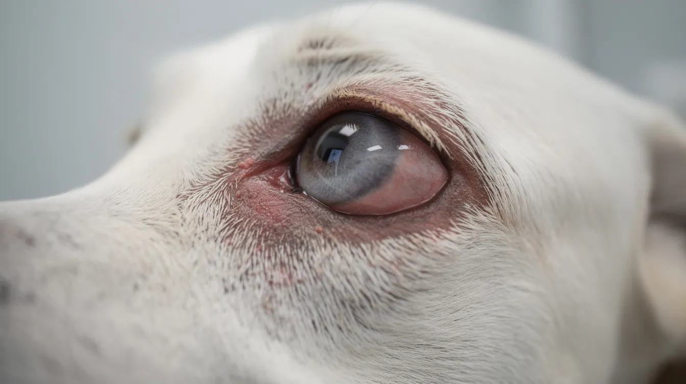
The earliest signs of glaucoma are often subtle signs that pet owners may initially attribute to minor irritation. Glaucoma may start with a slightly red eye. Dogs may squint due to glaucoma symptoms, show excessive tearing, or display mild episcleral redness in the affected eye. You might notice your dog showing reluctance to open one eye fully, sensitivity to bright light, or a faint cloudiness of the cornea that wasn’t there before.
Behavioral changes can also signal early trouble. A dog experiencing eye pain may become less active, hesitant to move in dim lighting, or show a decreased appetite. These common clinical signs are easy to dismiss, but in breeds predisposed to glaucoma, any persistent redness, squinting, or behavioral shift warrants prompt veterinary evaluation. If you’re unsure whether your dog is showing signs of illness versus normal aging, err on the side of caution with eye symptoms.
Advanced Symptoms
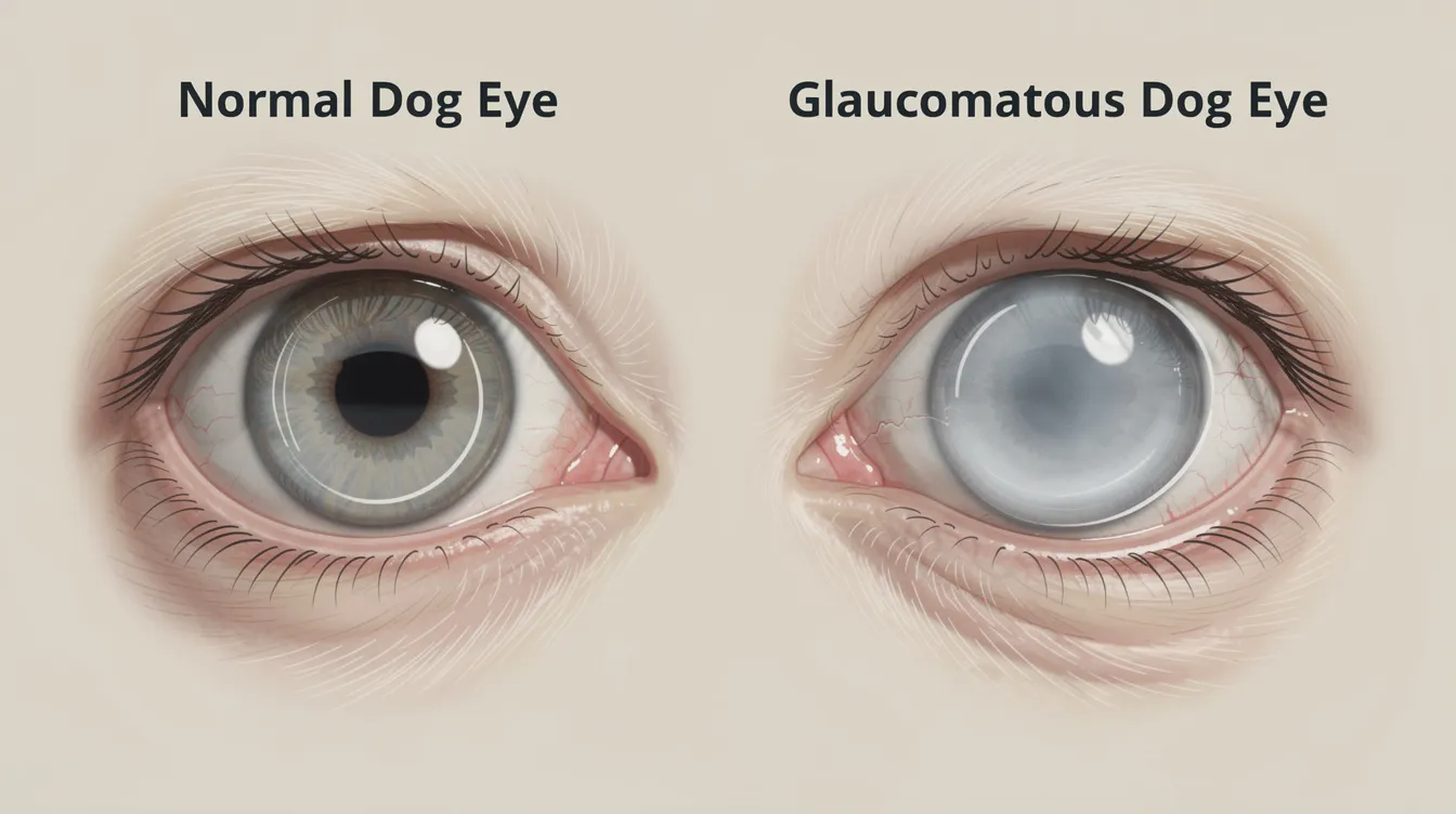
By the later stages of the disease, glaucoma symptoms become unmistakable. Symptoms indicating potential glaucoma include redness in the eye, a cloudy cornea, and visible pain. The cornea may take on a blue appearance due to edema. The eyeball may appear visibly enlarged (buphthalmos), and the pupil becomes a dilated pupil that doesn’t respond to light.
Glaucoma symptoms in dogs can include eye pain, cloudy or red eyes, bulging eyeballs, and vision loss. Pain behaviors become more pronounced – rubbing at the eye or head, lethargy, reluctance to be touched around the face, and reduced appetite. Complete vision loss manifests as bumping into furniture or walls, difficulty navigating familiar spaces, and startling easily. These advanced symptoms signal that significant, likely irreversible damage has already occurred, and emergency treatment is needed immediately to address whatever vision and comfort can still be salvaged.
Veterinary Diagnostic Procedures
Accurate diagnosis requires specific instrumentation and expertise. According to Cornell University College of Veterinary Medicine, proper diagnostic technique is essential for reliable results:
- Tonometry: A tonometer measures intraocular pressure IOP for diagnosis. Devices such as the TonoVet or TonoPen provide precise readings. Both eyes should be measured for accurate comparison, and proper technique – avoiding compression of the neck or globe – is critical. Normal canine IOP ranges from 20–28 mmHg; readings above 40–50 mmHg indicate a crisis requiring emergency intervention.
- Ophthalmoscopic examination: Diagnosis includes an internal eye examination with special instruments to evaluate the optic nerve head for cupping or degeneration, assess retinal health, and look for signs of lens displacement, uveitis, or tumors.
- Gonioscopy: This specialized test visualizes the drainage angle directly, helping classify whether glaucoma is primary or secondary and informing surgical candidacy. Missouri’s Veterinary Health Center emphasizes gonioscopy as particularly valuable for pre-surgical planning.
- Ocular ultrasound: When corneal opacity prevents direct visualization, ultrasound can assess lens position, detect intraocular tumors, and identify retinal detachment – all critical for determining the cause of the glaucoma and appropriate treatment.
Early diagnosis significantly improves prognosis for dogs with glaucoma, making routine screening especially important for predisposed breeds.
Treatment Options and Management
Treatment for glaucoma may involve medications to lower intraocular pressure or surgical options, depending on the severity, type, and whether useful vision remains. The primary goals are to reduce intraocular pressure to safe levels, preserve vision when possible, and ensure the dog’s comfort. Time is the most critical factor: every hour of uncontrolled pressure increases the risk of permanent damage.
Emergency Medical Treatment
When IOP exceeds 40–50 mmHg, emergency situations demand rapid pressure reduction. According to the Merck Veterinary Manual, emergency treatment typically follows this protocol:
- Topical prostaglandin analogs (e.g., latanoprost): Prostaglandin analogue eye drops can lower eye pressure rapidly by increasing uveoscleral outflow. However, the lens position must be assessed first – if an anteriorly luxated lens is present, the miosis these drugs induce can trap the lens and worsen pressure.
- Systemic osmotic diuretics: Mannitol (1–1.5 g/kg IV over 20–30 minutes) or glycerol (1–2 g/kg orally) rapidly dehydrates the vitreous to lower IOP. Water restriction for several hours after oral glycerol is standard.
- Topical and systemic carbonic anhydrase inhibitors: Dorzolamide or brinzolamide topically, or methazolamide systemically, reduce aqueous humor production by inhibiting carbonic anhydrase enzymes in the ciliary body.
- Beta blockers (e.g., timolol): Often used in combination with other agents to further reduce aqueous production.
Pressure reduction effects may begin within 15–30 minutes for prostaglandin analogs and osmotic agents, though sustained control often requires combination therapy. Referral to a veterinary ophthalmologist is recommended in most acute cases for specialized diagnostics and potential surgical intervention.
Long-term Medical Management
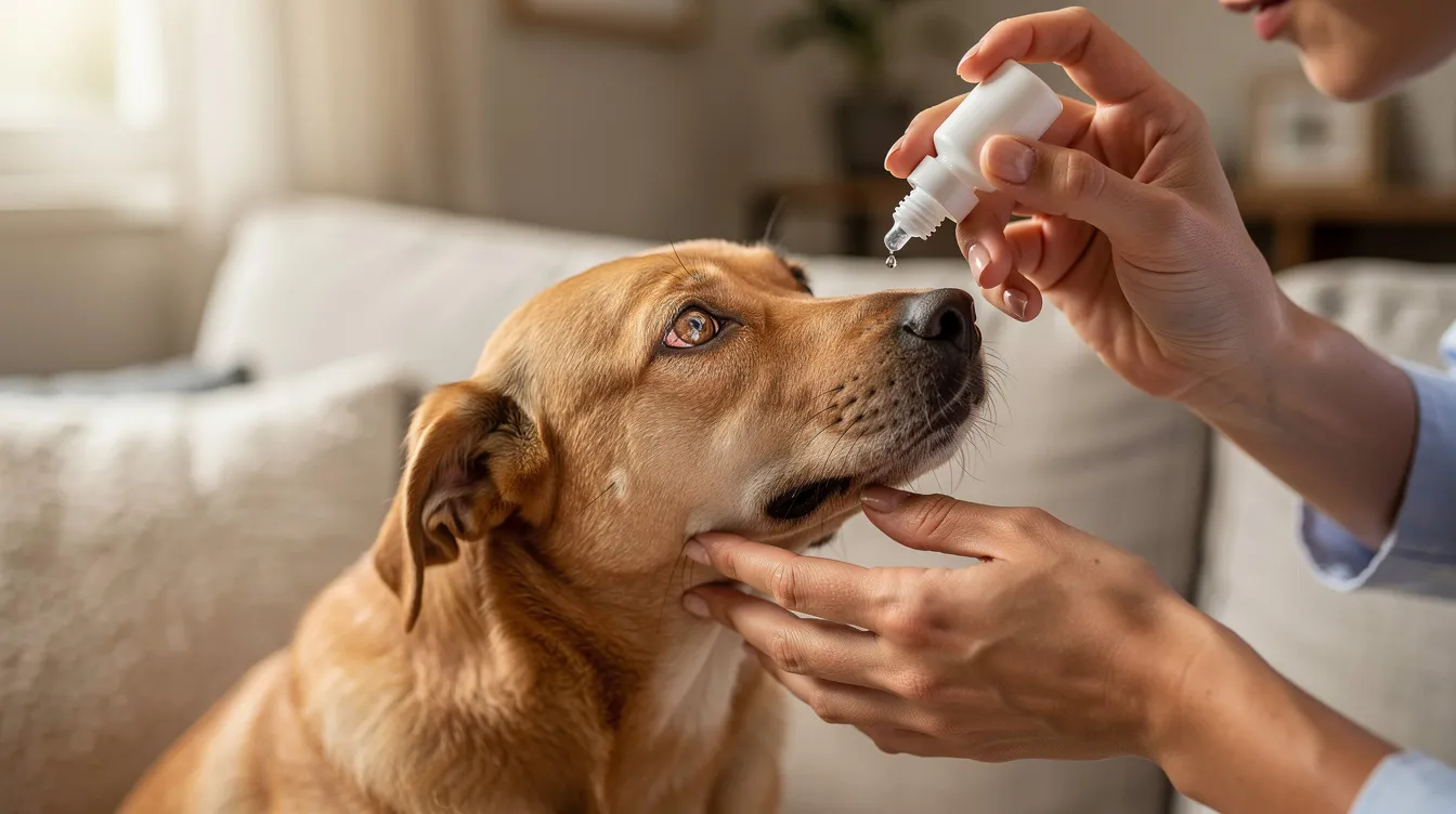
After stabilizing an acute crisis, long-term medical management typically involves daily eye drops administered on a strict schedule. Constant medical treatment is often required for long-term management:
- Prostaglandin analogs (latanoprost, travoprost, bimatoprost): Applied every 12–24 hours to maintain increased uveoscleral outflow. Contraindicated when lens luxation is present.
- Topical carbonic anhydrase inhibitors (dorzolamide 2%, brinzolamide 1%): Carbonic anhydrase inhibitors reduce fluid production in the eye. Applied every 8 hours, these are workhorses of chronic glaucoma management. Systemic formulations (methazolamide) are reserved for more severe cases.
- Beta blockers (timolol): Used as monotherapy in milder cases or combined with other medications. Dogs with cardiac conditions should be monitored for systemic side effects.
- Parasympathomimetic agents (pilocarpine, demecarium bromide): These increase conventional outflow through the drainage angle and may be used prophylactically in the unaffected eye when primary glaucoma has been confirmed in one eye.
Monitoring requires frequent IOP rechecks – every few days during the acute phase, transitioning to weekly and eventually monthly intervals as the disease stabilizes. Medication compliance is critical; missed doses frequently trigger pressure spikes and disease flare-ups. Combination drug formulations (e.g., dorzolamide/timolol) can reduce the number of daily drops, improving adherence.
Surgical Treatment Options
Surgical options may be necessary if medical treatment fails to maintain safe pressure levels, or when vision remains salvageable but unstable on medications alone. The choice among procedures depends on the type of glaucoma, remaining vision, and overall eye health.
| Procedure | Mechanism | Success/Outcomes | Key Considerations |
|---|---|---|---|
| Endoscopic cyclophotocoagulation (ECP) | Laser ablation of ciliary body epithelium to decrease aqueous production | IOP control in ~90% at 1 year, ~95% at 2 years; vision preserved in ~63% at 1 year | Complications include corneal ulcers (~28%), hypotony (~11%), retinal detachment (~11%) |
| Gonio-implantation (Ahmed valve) | Shunt device bypasses obstructed drainage angle | ~89% vision success at 1 year; mean vision preservation ~57.5 months | Requires management of granulation tissue around implant |
| Cyclocryotherapy | Freezing destruction of ciliary body to reduce aqueous production | ~83.6% success rate; mean IOP reduction of ~65% | Less invasive; may need repeat treatment |
| Enucleation/evisceration | Surgical removal of the eye or intraocular contents | Reliably eliminates pain; prosthetic option available | Reserved for irreversibly blind, painful eyes |
Enucleation may be necessary for irreversibly blind eyes where the goal shifts entirely to pain relief. Gentamicin injections can destroy the ciliary body to reduce pressure as a less invasive alternative to surgical removal, though this carries its own risk profile. Prognosis varies based on whether glaucoma is primary or secondary, and pre-operative vision status is the strongest predictor of post-surgical visual outcomes – dogs that are still visual at the time of surgery fare significantly better than those already blind in the affected eye.
Common Challenges and Solutions
Managing canine glaucoma long-term presents practical difficulties that go beyond the clinical aspects. Understanding these challenges upfront helps owners prepare and maintain consistent care.
Medication Administration Difficulties
Many owners find administering eye drops multiple times daily to be one of the hardest aspects of glaucoma management, especially when the dog is in pain or anxious about being touched near the eyes. Having your veterinary team demonstrate proper technique is invaluable. Approach from behind the dog’s head, tilt the nose slightly upward, and place drops into the lower conjunctival pocket rather than directly onto the cornea. Use treats and positive reinforcement to create a routine the dog can tolerate. Combination formulations that reduce the total number of daily applications can also make a meaningful difference in compliance.
Managing Treatment Costs
Glaucoma treatment costs add up substantially over time. Monthly medications may run several hundred dollars when multiple agents are required, regular IOP monitoring visits add ongoing expense, and surgical procedures like ECP or gonio-implantation can cost several thousand dollars depending on your region and hospital level. Planning ahead by discussing expected costs with your veterinarian, exploring pet insurance options (some policies cover glaucoma surgery, though coverage varies), and asking about generic medication alternatives can help manage the financial burden. Similar to managing other chronic conditions like dental disease or kidney failure, budgeting for ongoing care is essential.
Quality of Life Concerns
When vision loss does occur – whether partial or complete – dogs can still live fulfilling lives with appropriate environmental modifications. Keep furniture layouts consistent, use baby gates near stairs, and add scent markers or textured mats at doorways to help your dog navigate. Sound cues and verbal guidance become important communication tools. Pain management remains essential even after vision is gone; chronic glaucoma causes significant discomfort, and salvage procedures like enucleation often dramatically improve a dog’s quality of life by eliminating the source of constant pain. Many owners are surprised at how quickly their dog’s personality and energy return once a painful, blind eye is removed.
Conclusion and Next Steps
Glaucoma in dogs is a progressive eye disease where early intervention makes the decisive difference between preserving vision and losing it permanently. Understanding whether your dog faces primary or secondary glaucoma shapes every treatment decision, from emergency medications that reduce intraocular pressure to surgical procedures with documented success rates exceeding 80–90% for IOP control when performed in time.
Take these steps now:
- If your dog belongs to a high-risk breed, schedule a baseline eye pressure screening with your veterinarian – ideally including gonioscopy to evaluate drainage angle anatomy
- Learn to recognize the subtle signs: persistent redness, squinting, or behavioral changes in one eye warrant same-day veterinary evaluation
- If your dog has been diagnosed, discuss a comprehensive management plan including medication schedules, monitoring intervals, and criteria for surgical referral with a veterinary ophthalmologist
Long-term management expectations should be realistic: medical management often requires lifelong commitment, and many dogs will eventually need surgical intervention. However, with consistent care, many dogs maintain good quality of life for years. Keeping up with regular wellness examinations and monitoring for related eye conditions like eyelid tumors supports comprehensive ocular health.
Note for cat owners: while this guide focuses on dogs, glaucoma in cats also occurs. Feline glaucoma shares some similarities but differs in key ways – secondary glaucoma from anterior uveitis is the most common form in most cats, and certain breeds like Burmese and Siamese cats may have higher susceptibility. If you suspect signs of glaucoma in your cat, seek veterinary evaluation promptly.
Frequently Asked Questions
Can dogs live normal lives with glaucoma?
Many dogs with glaucoma maintain good quality of life, especially with early detection and aggressive treatment. While vision loss is common in most cases, dogs adapt remarkably well to reduced or absent sight when their pain is controlled. Even dogs that lose both eyes can navigate familiar environments, play, and enjoy daily life when owners provide consistent environmental support.
How quickly can glaucoma cause blindness in dogs?
In acute glaucoma with very high IOP, retinal and optic nerve damage can begin within hours. Blindness can occur if glaucoma is untreated for too long – sometimes within the same day during an acute crisis. Chronic glaucoma progresses more gradually, with cumulative damage causing vision loss over weeks to months. This is why any sudden eye redness, pain, or cloudiness should be treated as an emergency.
Is glaucoma painful for dogs?
Yes, significantly. Increased pressure within the eye causes pain comparable to severe headaches in humans. Dogs may show pain through squinting, rubbing at the affected eye, lethargy, decreased appetite, and reluctance to play or be active. Even after vision is lost, many dogs remain in considerable discomfort until the source of increased IOP is addressed through medical treatment or surgery.
What happens if my dog’s glaucoma is left untreated?
Without treatment, glaucoma causes irreversible blindness, structural damage to the eye including optic nerve death and retinal atrophy, progressive enlargement of the globe (buphthalmos), and ongoing pain. The eye may eventually require surgical removal as the only humane option. In some cases, chronic inflammation from an untreated glaucomatous eye can also affect overall wellbeing.
How much does glaucoma treatment cost?
Costs vary by region and treatment level. Daily medical management with multiple eye drops may cost several hundred dollars monthly. Surgical procedures such as endoscopic cyclophotocoagulation or gonio-implantation typically cost several thousand dollars per eye. Ongoing monitoring visits add further expense. Discussing a financial plan with your veterinarian early helps avoid difficult decisions later in the disease course.
Should both eyes be treated if only one shows signs of glaucoma?
In primary glaucoma, absolutely. Because the underlying anatomical abnormality is bilateral, the unaffected eye carries very high risk of developing glaucoma – often within months. Prophylactic therapy with medications such as demecarium bromide every 24 hours or timolol every 12 hours, sometimes combined with anti-inflammatory agents, may delay onset in the second eye. Your veterinary ophthalmologist should evaluate and monitor both eyes even when only one eye currently shows elevated pressure.
Can glaucoma in dogs be prevented?
Primary glaucoma cannot be fully prevented because it stems from inherited anatomy, but risk can be mitigated through breed screening programs, avoiding breeding dogs with glaucoma history, and baseline gonioscopy in predisposed breeds. Secondary glaucoma prevention focuses on prompt treatment of eye injuries, uveitis, lens luxation, and other underlying conditions before they compromise drainage. Regular veterinary eye examinations remain the best tool for early detection.
When should I consider eye removal for my dog’s glaucoma?
Enucleation or evisceration should be considered when vision in the affected eye is confirmed irreversibly lost and the eye remains painful despite medical and surgical management. Many owners are initially resistant to the idea, but dogs that undergo eye removal for end-stage glaucoma typically show dramatic improvements in comfort, energy, and overall quality of life within days of surgery. The decision is ultimately about restoring comfort rather than appearance.The decision is ultimately about restoring comfort rather than appearance.
Article reviewed by Dr. Roger Hart, DVM
Leave a Reply