Introduction
Otitis media symptoms in dogs and cats signal a middle ear infection that demands prompt attention-these signs range from subtle head shaking and ear pain to serious neurological issues like loss of balance, facial nerve paralysis, and vestibular disease. This comprehensive guide covers how to recognize every stage of otitis media symptoms in companion animals, including otitis media vestibular sydrome. Furthermore, understanding the implications of otitis media vestibular sydrome is crucial for early diagnosis and effective treatment. It is important for pet owners to be aware of the signs and symptoms of otitis media vestibular sydrome to ensure their pets receive prompt care.
This content focuses specifically on middle ear infection symptoms in dogs and cats-not outer ear infections (otitis externa) alone, nor human ear conditions. It is written for pet owners who want to understand what their animal may be experiencing, assess symptom severity, and make informed decisions about seeking veterinary attention. Otitis media is a middle ear infection caused by bacteria or viruses, and in companion animals, it often develops as a secondary complication of chronic ear infections or upper respiratory disease. Because the middle ear sits adjacent to the inner ear and the vestibular system responsible for maintaining balance, untreated infections can progress rapidly toward permanent damage, including conditions like otitis media vestibular sydrome. Recognizing the signs of otitis media vestibular sydrome can help ensure timely intervention.
In addition, the frequency of otitis media vestibular sydrome cases has increased, making it an important topic for pet owners to understand. Being educated about otitis media vestibular sydrome will help you recognize the signs early and seek veterinary assistance.
Moreover, if your pet shows any symptoms related to otitis media vestibular sydrome, it is essential to consult your veterinarian as soon as possible to mitigate potential complications of the condition.
Pet owners should be aware that otitis media vestibular sydrome can lead to more severe neurological symptoms if not treated promptly. Regular check-ups with a veterinarian can help monitor for any signs of this condition.
Recognizing the signs and symptoms of otitis media vestibular sydrome can significantly influence your pet’s health outcomes. Early intervention is key to managing this condition.
Consult your veterinarian to discuss the potential implications of otitis media vestibular sydrome on your pet’s overall health.
The most common otitis media symptoms include head tilt, ear pain when the base of the ear or jaw is touched, head shaking, discharge with foul odor, and balance disturbances. Fluid build-up in the middle ear leads to inflammation and pain, and when infection spreads to the inner ear, dogs and cats may show sudden loss of balance and disorientation.
By reading this guide, you will:
- Learn to identify early warning signs before symptoms become severe
- Understand the difference between mild, moderate, and severe otitis media presentations
- Know exactly when to seek urgent care or emergency veterinary attention
- Differentiate otitis media symptoms from other ear problems and conditions
- Support your pet’s recovery with informed observation and communication with your veterinarian
Reviewed by Dr. Roger Hart, DVM for accuracy.
Understanding Otitis Media in Companion Animals
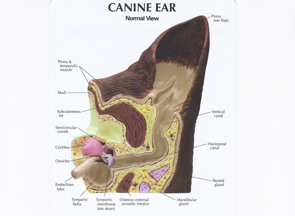
Otitis media refers to inflammation or infection of the middle ear-the space behind the tympanic membrane (eardrum) that houses the tympanic cavity, auditory ossicles, and tympanic bulla. Understanding what happens in this part of the ear helps pet owners recognize why symptoms extend far beyond simple ear discomfort and can affect a dog’s balance, nervous system function, and daily life.
What is Otitis Media and Inner Ear Involvement
The middle ear is a small, air-filled chamber separated from the outer ear canal by the eardrum. In dogs and cats, this space connects to the throat via the auditory (Eustachian) tube and sits immediately adjacent to the inner ear, where the vestibular system and hearing organs reside. When bacteria, yeast, or inflammatory fluid accumulates behind the eardrum, infection develops in this confined space and can place pressure on surrounding structures.
Otitis media differs significantly from otitis externa (outer ear infections). While otitis externa involves the visible ear canal and produces familiar signs like redness, odor, and scratching, otitis media affects deeper structures that cannot be seen without specialized equipment. According to the Merck Veterinary Manual, otitis media often arises secondarily to chronic otitis externa or through extension via the auditory tube, and many cases in dogs are associated with chronic ear canal disease.
The proximity of the middle ear to the vestibular system explains why middle ear infections can cause vestibular disease in dogs-producing head tilt, nystagmus (rapid eye movement), and ataxia that mimic stroke or brain disease. The vestibular system includes peripheral ear structures in the inner and middle ear, while its central components located in the brain help maintain balance and spatial orientation. Inner ear problems may trigger vestibular disease in older dogs, making the distinction between middle and inner ear involvement clinically important.
In cats, the tympanic cavity has a unique anatomical feature: a bony septum divides it into ventromedial and dorsolateral compartments that communicate only through a small dorsal gap. This anatomy affects how fluid or infection distributes within the feline middle ear and can complicate both diagnosis and treatment.
Types of Otitis Media
Understanding the relationship between ear infections and otitis media vestibular sydrome is vital for pet owners. This connection can help in identifying when medical attention is needed.
Acute versus chronic otitis media present differently and carry different implications. Acute cases develop over hours to days with prominent signs-sudden onset of head tilt, pain, and possible vestibular symptoms. Chronic otitis media may show subtle or recurring clinical signs, with structural changes like thickened tympanic bullae or bone proliferation developing over time.
Primary versus secondary infections also differ in their underlying cause:
- Primary otitis media develops without preceding outer ear disease. A notable example is primary secretory otitis media (PSOM), especially seen in Cavalier King Charles Spaniels, where mucus accumulates in the middle ear due to Eustachian tube dysfunction.
- Secondary otitis media stems from other causes-chronic otitis externa, allergies, foreign bodies, nasopharyngeal polyps (particularly common in cats), parasitic infestation, or upper respiratory disease.
Untreated chronic external ear infections remain one of the most common pathways to middle ear involvement. This relationship between outer and inner and middle ear disease underscores why recurring ear infections should never be dismissed as routine. If your dog is shaking its head repeatedly, it may indicate more than a simple outer ear problem.
Understanding these types leads directly to recognizing the specific symptoms each presentation produces.
Recognizing Otitis Media Symptoms and Clinical Signs
With a foundation in what otitis media is and how it develops, identifying the specific symptoms becomes the critical next step. Symptoms range from barely noticeable behavioral shifts to dramatic neurological signs, and recognizing early changes can prevent progression to severe disease. Common symptoms of otitis media include ear pain and fever, though the presentation in animals requires careful observation since pets cannot verbalize their discomfort.
Early Warning Signs
Be proactive about checking for signs of otitis media vestibular sydrome, especially if your pet shows any unusual behavior.
The earliest otitis media symptoms are often easy to overlook, particularly in cats. Watch for:
- Occasional head shaking or ear scratching: Your pet may paw at or rub the affected ear against furniture or the floor. This behavior may appear intermittent and inconsistent. Tugging at the ear is a classic sign of ear irritation-and while this description comes from pediatric medicine, the equivalent behavior in pets (scratching, pawing, rubbing) serves the same purpose.
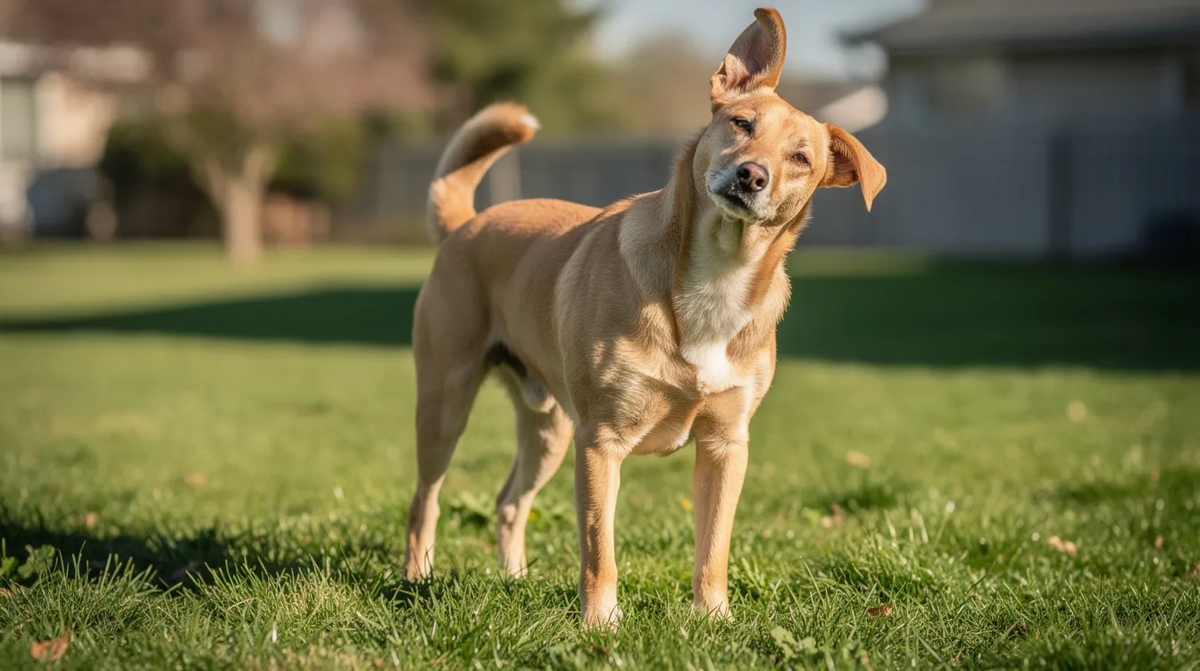
- Mild head tilt: A subtle, intermittent tilt of the head position toward one side, sometimes noticed only during rest or when your pet is listening. Even a mild head tilt warrants monitoring.
- Changes in hearing responsiveness: Your pet may fail to respond to commands, sounds, or their name-suggesting possible hearing impairment. Ear infections can cause reduced hearing or difficulty hearing, and this diminished responsiveness is frequently among the first signs pet owners notice.
- Mild ear changes: Slight redness or warmth at the ear pinna, increased wax or cerumen production, or a faint odor from the ear canal may indicate early infection.
These early signs frequently overlap with simple otitis externa, making differentiation challenging without veterinary examination. Many dogs with early middle ear involvement continue eating normally and show no balance disturbances, which delays recognition.
It’s essential to keep an eye out for early signs of otitis media vestibular sydrome, particularly if your pet displays behavioral changes or unusual movements.
Advanced Symptoms
When infection progresses deeper into the middle ear or begins affecting adjacent structures, symptoms become more pronounced and concerning:
- Persistent head tilt: A consistent, noticeable tilt toward the affected side that does not resolve. Dogs may lean or fall in the direction of their head tilt, and this loss of balance can worsen with movement.
- Pain responses: Significant discomfort when the ear base is touched, when opening the jaw, or when the submandibular region is palpated. Some animals show aggression or withdrawal when the ear area is approached. It’s important to seek medical attention if symptoms last more than 2 to 3 days, as persistent pain suggests deeper involvement. Unrelenting severe pain requires immediate medical care.
- Ear discharge and odor: Purulent (pus-like), brown, or yellow discharge from the ear canal, often accompanied by a strong foul odor. Fluid or pus drainage may indicate a ruptured eardrum, which requires prompt veterinary evaluation. Swelling or visible obstruction of the canal may also develop.
- Balance and coordination problems: Ataxia or incoordination-stumbling, circling, or inability to walk in a straight line. In cats, difficulty jumping or navigating stairs may become obvious. These signs indicate involvement of the vestibular system and potential progression toward vestibular disease.
Dogs may show sudden loss of balance and disorientation, and common symptoms include head tilt and irregular eye movements. Nystagmus is characterized by irregular, jerking eye movements-a hallmark sign of vestibular involvement that often accompanies advanced otitis media. When this eye movements pattern appears, the infection has likely reached or is affecting the inner ear.
Signs of facial weakness can indicate complications from ear infections. Facial nerve paralysis-drooping of the lip, facial asymmetry, inability to blink, or ear drooping on the affected side-occurs because the facial nerve passes through the middle ear. Horner syndrome (sunken eye, constricted pupil, drooping eyelid) on the same side as the affected ear represents another serious neurological complication.
Behavioral Changes
Beyond physical symptoms, otitis media produces notable behavioral changes that pet owners should monitor:
- Eating difficulties: Reluctance to eat or difficulty chewing, particularly on the affected side, because jaw movement intensifies middle ear pain. A dog not eating and showing lethargy or a cat refusing food alongside ear symptoms should raise concern. Difficulty sleeping may worsen in animals with ear infections, as lying on the affected side increases pressure and pain.
- Social withdrawal: Decreased activity, reluctance to play, hiding (especially in cats), and avoidance of interaction. Excessive crying or fussiness can indicate ear infection in young animals, just as it does in young children.
- Restlessness and vomiting reluctance: Disrupted sleep patterns, pacing, or inability to settle. Some pets experience nausea or motion sickness due to vestibular involvement, showing vomiting or drooling alongside experiencing dizziness.
Key symptom progression summary: Otitis media typically begins with intermittent scratching and head shaking, progresses to persistent head tilt and pain, and can advance to vestibular signs, facial paralysis, and hearing loss. Signs typically do not progress or worsen over time once appropriate treatment begins-but without intervention, the trajectory is toward increasingly severe disease.
The transition from recognizing symptoms to accurately assessing their severity determines how urgently your pet needs professional evaluation.

Detailed Symptom Assessment and Veterinary Evaluation
Professional diagnostic testing is essential for confirming otitis media because external examination alone cannot reliably identify middle ear involvement. Building on the symptom categories above, this section provides tools for structured observation and a framework for assessing urgency.
Pet Owner Observation Checklist
As the condition progresses, otitis media vestibular sydrome can lead to significant health issues. Recognizing symptoms early can prevent complications.
Veterinary advice is essential to manage otitis media vestibular sydrome effectively. Always seek professional assistance when symptoms arise.
Before your veterinary visit, systematically document what you’re observing. This medical history helps your veterinarian determine the appropriate physical exam approach and whether advanced imaging like a CT scan is necessary:
- Monitor head position and movement: Note whether the head tilt is constant or intermittent, which direction it favors, and whether your pet circles or falls. Record any eye movements (nystagmus) you observe.
- Check for ear discharge or odor: Examine both ears visually (without inserting anything into the canal). Note the color, consistency, and smell of any discharge. Compare both ears for asymmetry in redness or swelling.
- Assess eating and drinking behavior: Track whether your pet is eating normally, favoring one side while chewing, or refusing food entirely. Note if drinking water has become difficult or messy.
- Note balance and coordination changes: Observe your pet walking, turning, and navigating stairs or furniture. Document stumbling, falling, or reluctance to move. In cats, note any changes in jumping ability or grooming habits.
Symptom Severity and Underlying Cause Comparison
Symptom Category | Mild | Moderate | Severe |
|---|---|---|---|
Head Tilt | Occasional, intermittent tilting | Consistent tilt to one side | Persistent tilt with circling or falling |
Pain Indicators | Mild discomfort when ear touched | Withdrawal or vocalization on palpation | Severe pain; aggression when ear or jaw area approached |
Balance Issues | No observable imbalance | Occasional stumbling or leaning | Ataxia, falling, inability to stand; vestibular disease signs |
Eating/Drinking | Normal appetite | Reluctance to chew hard food | Refusal to eat; weight loss |
Discharge | Minimal or mild odor | Visible discharge; moderate odor | Purulent drainage; severe odor; possible ruptured eardrum |
Neurological Signs | None | Mild facial asymmetry or Horner signs beginning | Facial paralysis; nystagmus; Horner syndrome |
Interpreting severity: Mild symptoms warrant a scheduled veterinary appointment within days. Moderate symptoms call for prompt evaluation-ideally within 24 to 48 hours. Severe symptoms, particularly sudden onset vestibular signs, facial nerve paralysis, or refusal to eat, require urgent care or emergency room evaluation.
Veterinary diagnostic evaluation typically includes otoscopic examination of the tympanic membrane (checking whether it is intact, bulging, opaque, or ruptured), culture and cytology of any accessible discharge, and advanced imaging. When vestibular signs are present, a broader workup may also include blood pressure measurement to assess overall health and rule out contributing conditions. CT scan or MRI is particularly valuable for assessing the tympanic bulla, detecting fluid accumulation, wall thickening, and bone changes that indicate chronic disease. In a study of 16 cats with diagnosed otitis media, CT/MRI findings showed fluid in tympanic bullae in nearly all cases, confirming that imaging often reveals disease invisible to external examination.
In a study of 26 cats with suppurative otitis media and intact tympanic membranes, neurological signs were the most common presentation (15 out of 26 cats), followed by otalgia (ear pain) in 9 out of 26, while external ear discharge appeared in only 5 out of 26. This data underscores that otitis media can exist with minimal external signs-diagnostic testing beyond surface-level examination is frequently necessary.
Common Challenges, Treatment Options, and Solutions
Recognizing otitis media symptoms presents several factors that complicate accurate identification, particularly for pet owners without veterinary training.
Hidden Symptoms in Cats
Cats are exceptional at masking illness. According to the MSD Veterinary Manual, early otitis media signs in cats are often unnoticed until the disease has progressed significantly. A cat may stop grooming one ear, hold its head position slightly differently, or simply become quieter-changes easily attributed to mood rather than disease.
Solution: Establish baseline behavioral awareness. Know your cat’s normal ear position, grooming patterns, activity level, and appetite. Any deviation from baseline lasting more than 24 to 48 hours warrants closer observation. In a study of 196 cats with peripheral vestibular syndrome, otitis media/interna was the most common cause, and a history of otitis externa or upper respiratory signs, facial nerve paralysis, and Horner syndrome strongly predicted middle ear involvement.
Recurring Infections
Many dogs and cats experience repeated episodes of ear disease. Chronic or incompletely treated otitis externa can silently progress to middle ear involvement. In dogs with peripheral vestibular disease (n = 188), otitis media/interna was diagnosed in approximately 49 cases (about 26%), making it a leading identified cause of vestibular symptoms after idiopathic vestibular syndrome.
Solution: Complete every prescribed treatment course fully-typical medical therapy for feline otitis media lasts 3 to 6 weeks. Follow up with your veterinarian even if symptoms appear resolved. Chronic cases are more likely to produce structural changes and irreversible nerve damage, while early treatment improves prognosis dramatically. Certain medications can be toxic to the inner ear, so always use ear medications only as prescribed.
Misidentifying Symptoms
Otitis media symptoms overlap significantly with other causes of vestibular disease, including idiopathic vestibular syndrome (particularly old dog vestibular disease in senior dogs), brain tumors causing central vestibular disease, head trauma, hypothyroidism, and stroke. Hypothyroidism is a potential cause of vestibular disease, and trauma or injury can lead to vestibular disease in dogs. The clinical signs can appear identical to the untrained eye.
Solution: Never attempt to diagnose or treat suspected middle ear disease at home. A veterinary physical exam combined with diagnostic testing-including imaging and potentially blood work-is required to identify the specific cause. Brain tumors may cause central vestibular disease in dogs, and the treatment plan differs dramatically from that of otitis media. Professional evaluation ensures your pet receives appropriate treatment rather than delayed or incorrect intervention, as dental pain can also cause similar reluctance to eat and facial sensitivity.
If surgical intervention becomes necessary for refractory cases, understanding what soft tissue surgery entails can help pet owners prepare.
Conclusion and Next Steps
Discussing your pet’s symptoms related to otitis media vestibular sydrome with your veterinarian is crucial for accurate diagnosis and treatment planning.
Otitis media symptoms in dogs and cats span a wide spectrum-from occasional head shaking and subtle behavioral changes to severe vestibular disease, facial nerve paralysis, and hearing loss. The critical takeaway is that middle ear infections often present with minimal external signs while causing significant internal disease, and early recognition paired with professional veterinary care offers the best outcomes. Most dogs recover from vestibular disease within two to three weeks when the underlying condition is properly addressed, and many dogs improve within two to three weeks without treatment in idiopathic cases. Supportive care often resolves vestibular disease in dogs, though some dogs may retain a mild head tilt for life.
Additionally, inform your vet if you suspect your pet may be experiencing symptoms associated with otitis media vestibular sydrome.
Immediate steps if you suspect otitis media:
- Schedule a veterinary examination promptly-within 24 to 48 hours for moderate symptoms, or seek emergency care for sudden vestibular signs, refusal to eat, or facial paralysis
- Document symptoms and behaviors using the observation checklist: note timing, progression, affected side, and any triggers
- Prepare questions for your veterinary visit: ask about diagnostic testing options, whether imaging is recommended, the expected treatment plan and duration, and signs that would indicate the need for emergency room evaluation
Most dogs improve within 72 hours of diagnosis and appropriate treatment. Complete recovery often occurs within 2 to 3 weeks. Dogs may require hospitalization for severe vestibular disease cases, where supportive care, nursing care, anti nausea medications, and intravenous fluids can be administered. Physical therapy may also be recommended during recovery to help rebuild coordination and muscle strength in pets with persistent balance deficits. Medications for nausea can aid in vestibular disease treatment, while corticosteroids are generally not recommended for vestibular disease.
Related topics worth exploring include preventive ear cleaning techniques, management of allergies as an underlying cause of chronic ear disease, and long-term treatment options for pets with recurrent ear problems.
Professional Consultation and Resources
If your pet is showing any signs discussed in this guide-particularly head tilt, loss of balance, ear discharge, or pain-professional veterinary evaluation should be your first step. Dr. Roger Hart, DVM, offers specialized care for companion animals experiencing ear problems and vestibular symptoms, with expertise in both diagnostic evaluation and comprehensive treatment plans for middle ear disease.
For additional reading on otitis media and vestibular disease in companion animals, the following resources provide authoritative veterinary information:
- Merck Veterinary Manual: Otitis Media and Interna in Animals – comprehensive clinical reference covering causes, signs, diagnosis, and treatment
- Cornell University College of Veterinary Medicine – evidence-based resources on feline and canine health conditions
Awareness of otitis media vestibular sydrome can significantly impact the care and treatment your pet receives.
This article has been reviewed by Dr. Roger Hart, DVM for accuracy.
Frequently Asked Questions
Be vigilant for any signs of otitis media vestibular sydrome, especially during seasonal changes when ear infections can be more prevalent.
Understanding the nuances of otitis media vestibular sydrome helps in differentiating it from other conditions that might cause similar symptoms.
Can otitis media symptoms appear suddenly in healthy pets?
Yes-many cats and dogs present acutely (within 48 hours) with dramatic neurological signs including head tilt, nystagmus, and ataxia, even when their medical history does not include obvious prior ear disease. However, investigation often reveals a background of subclinical outer ear disease or upper respiratory infection that went undetected. Idiopathic cases often resolve without treatment in 2 to 3 weeks, but sudden onset vestibular signs always warrant veterinary evaluation to rule out serious other causes including brain tumors or stroke.
How do I differentiate between otitis media and simple ear wax buildup?
Simple cerumen (ear wax) buildup produces mild discharge without significant pain, no neurological signs, and no systemic symptoms. Otitis media is distinguished by the presence of head tilt, nystagmus or other eye movements abnormalities, cranial nerve deficits such as facial paralysis, pain when touching the ear base or opening the jaw, and balance disturbances. Definitive differentiation requires veterinary examination and often imaging to detect fluid or structural changes in the tympanic bulla.
Are certain dog or cat breeds more prone to otitis media symptoms?
In dogs, Cavalier King Charles Spaniels are predisposed to primary secretory otitis media (PSOM). Breeds with pendulous ears, narrow ear canals, or predisposition to allergies (such as Cocker Spaniels, Basset Hounds, and Labrador Retrievers) face higher risk due to increased susceptibility to chronic otitis externa, which can progress to middle ear involvement. In cats, domestic shorthairs are the most commonly reported breed in otitis media studies, though no single feline breed shows strong predisposition. Cats with nasopharyngeal polyps are at particular risk.
What should I do if symptoms appear during evening or weekend hours?
If your pet shows sudden onset severe vestibular signs (inability to stand, falling, continuous nystagmus), severe pain, facial paralysis, or refusal to eat or drink, seek emergency care at an after-hours veterinary facility. Mild symptoms such as occasional head shaking or scratching can generally wait for a regular veterinary appointment the next business day. A high fever in infants may require urgent medical evaluation-similarly, severe neurological signs in pets should not wait.
Understanding the potential for otitis media vestibular sydrome can help you monitor your pet more effectively and seek the necessary veterinary care.
How long do otitis media symptoms typically last with treatment?
Otitis media vestibular sydrome can complicate recovery; therefore, early recognition and treatment are essential.
Most patients show improvement within 72 hours of beginning appropriate treatment. Complete recovery often occurs within 2 to 3 weeks, though treatment courses for middle ear infections typically last 3 to 6 weeks to ensure full resolution. Small tympanic membrane perforations may heal in 2 to 3 weeks. Some dogs may retain a mild head tilt permanently, but signs typically do not progress or worsen over time once treatment is underway. Even after successful treatment, some neurological signs such as persistent head tilt or facial nerve paralysis may remain, particularly in cats with inner ear involvement.
Can otitis media symptoms indicate other underlying health problems?
Absolutely. Middle ear infections frequently develop secondary to an underlying condition-chronic allergies, hypothyroidism, autoimmune disease, nasopharyngeal polyps, or upper respiratory infections can all predispose pets to otitis media. Additionally, the neurological symptoms of otitis media (head tilt, nystagmus, ataxia) overlap with several factors including idiopathic vestibular syndrome, central vestibular disease from brain tumors, head trauma effects, and other neurological issues. Proper diagnostic testing identifies whether otitis media is the primary problem or a manifestation of broader disease, ensuring the treatment plan addresses the true underlying cause.
Be sure to inform your veterinarian if you suspect your pet may be suffering from otitis media vestibular sydrome, as this information can guide their assessment and treatment strategy.
Discuss with your veterinarian about how to prevent otitis media vestibular sydrome in breeds that are predisposed to ear issues.
Research shows that prompt treatment of otitis media vestibular sydrome can lead to better outcomes in affected pets.
Understanding the risks associated with otitis media vestibular sydrome can help pet owners take preventative measures.
Be proactive in recognizing the signs of otitis media vestibular sydrome. Early detection is key to effective management and treatment.
Prompt recognition of otitis media vestibular sydrome is critical for ensuring that pets receive timely treatment to prevent long-term complications.
Understanding the various symptoms and implications of otitis media vestibular sydrome will empower pet owners to make informed decisions regarding their pet’s health.
Leave a Reply