
Introduction
Bumps on dogs skin can range from harmless to serious growths, and discovering an unexpected lump on your dog’s skin naturally triggers concern. This visual guide provides detailed descriptions and photographic references of the most common skin bumps on dogs skin, helping pet parents compare what they see at home with known benign and malignant growths.
This guide covers the full spectrum of skin lumps-from soft, harmless lipomas to aggressive mast cell tumors-with emphasis on visual characteristics, typical locations on a dog’s body, and growth patterns. While pictures help with initial identification, only veterinary examination with tools like fine needle aspiration and biopsy can provide an accurate diagnosis and appropriate treatment plan. Various benign and malignant lumps, including bumps on dogs skin, can appear similar, making professional evaluation essential.
Understanding bumps on dogs skin is crucial for early detection and treatment. Various types of lumps can occur, and they often vary in appearance and significance.
This resource is designed for dog owners who have discovered a new bump and need a visual reference to understand what they might be dealing with before their veterinary appointment.
By the end of this guide, you will be able to:
- Visually distinguish common benign growths from potentially cancerous tumors
- Recognize red flag features that require immediate veterinary attention
- Perform a safe at-home examination and document findings for your vet
- Understand which dog breeds and age groups are most susceptible to specific skin bumps
- Know when home monitoring is acceptable versus when professional care is urgent
Understanding Bumps on Dogs
A bump or skin growth on a dog refers to any visible or palpable abnormal tissue mass on or beneath the skin surface. These skin lumps vary enormously in size-from a tiny small bump just a few millimeters across to masses several centimeters wide. Textures range from soft and squishy to rock-firm. Colors span skin-tone, pink, red, brown, and black. Some are freely movable beneath the skin, while firm, fixed masses that do not move under the skin may indicate a problem.
Dogs develop skin bumps for many reasons. Normal aging causes growths like lipomas and skin tags to appear more frequently. Viral infections trigger papillomas. Blocked oil glands and hair follicles produce cysts. Cellular growth regulation failures lead to neoplasms-both benign and malignant. Sun exposure can contribute to squamous cell carcinomas, hormonal imbalances affect glandular tumors, and immune system responses play a role in histiocytomas. The tissue of origin-whether fat cells, sebaceous glands, skin cells, connective tissue, blood vessels, or pigment cells-determines the type of growth.
Benign vs Malignant Growths
Regular inspections of bumps on dogs skin help in early identification of potential problems.
Benign growths are non-cancerous masses that do not invade surrounding normal tissue or spread to other organs. Common examples include lipomas (soft fatty tumors), papillomas (viral warts), histiocytomas (button tumors in young dogs), sebaceous cysts, and skin tags. Visually, these tend to be slow-growing, soft or moderately firm, freely movable under the skin, and often painless. Many benign lumps can include lipomas and sebaceous cysts that remain stable for months or even years.
Knowing more about bumps on dogs skin can help you take proactive steps to ensure your pet’s health.
Malignant tumors can invade tissue and spread to organs, making early identification critical. Cancerous tumors such as mast cell tumors, malignant melanomas, squamous cell carcinomas, and hemangiosarcomas tend to share certain visual warning signs: rapid growth over weeks rather than months, irregular or poorly defined borders, ulceration or bleeding, color changes, and fixation to deeper tissues. Mast cell tumors are the most common malignant tumors in dogs, and they are notoriously difficult to identify visually because they mimic many other skin conditions.
When examining bumps on dogs skin, it’s essential to be aware of their characteristics to identify which require immediate veterinary attention.
Normal vs Concerning Features
Pet parents sometimes mistake normal skin variations for abnormal bumps. Nipples (present on both male dogs and females), skin folds common to certain dog breeds, calloused areas on elbows and joints, and flat pigmentation spots are all normal tissue features that can cause unnecessary alarm.
Veterinarians use specific signs to determine if a growth needs examination. Red flag features include: rapid growth of a lump on a dog, ulceration or bleeding from the skin surface, sudden pigment changes, hair loss and missing fur around the lesion, swelling that increases and decreases (characteristic of mast cell tumors releasing histamine), firm attachment to deeper tissues, irregular shapes or borders, and pain or warmth when touched. Lumps in sensitive areas-such as the mouth, nail beds, or near joints-need immediate veterinary attention.
Common Benign Bumps with Pictures
Many lumps on dogs are benign and common, particularly in older dogs, and benign dog lumps commonly include lipomas and sebaceous cysts. However, even obviously harmless-looking growths should be monitored for changes in size, color, texture, or behavior. The following sections describe the most frequently encountered benign growths with visual details to aid identification.
Papillomas (Warts)
Monitoring bumps on dogs skin is particularly important as they can change over time, leading to more serious conditions.
Many dogs with bumps on dogs skin exhibit no signs of discomfort, but it’s crucial to observe their behavior closely.
Papillomas are contagious warts often found in young dogs, caused by canine papillomavirus. Their visual hallmark is a small, fleshy, cauliflower-like growth that may appear white, pink, or slightly scaly. Papillomas are contagious and often found in young dogs, typically appearing around the mouth, lips, and gums in dogs under 2 years old. Older dogs may develop them near eyelids or on the dog’s head.
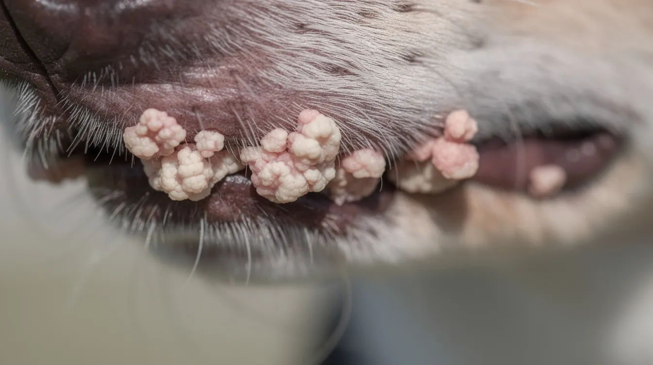
Papillomas typically range from a few millimeters to about a centimeter across. Early-stage warts may look like tiny smooth bumps before developing their characteristic rough, textured surface. In young dogs, the immune system usually clears papillomas within 1–3 months without treatment. Severe or persistent cases may require surgical removal or cryotherapy.
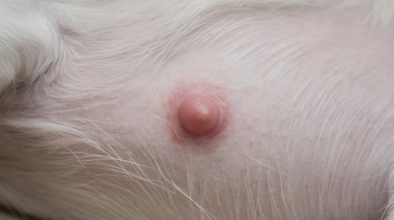
Sebaceous Cysts
Sebaceous cysts result from blockage of sebaceous glands or hair follicles beneath the dog’s coat. They present as firm, round lumps under the skin with a smooth surface-often described as a round lump that feels like a marble or pea beneath the skin. These cysts fill with keratin and sebum, ranging from pea-sized to golf-ball-sized.
When intact, cysts feel firm and well-defined. If ruptured or infected, they may release a thick, waxy, or cheesy material and develop redness consistent with a bacterial infection. Follicular cysts are benign bumps that can become itchy or painful as they grow larger. Dog breeds with oily or dense coats are particularly prone to developing these cysts. Treatment consists of monitoring for small, stable cysts; larger or infected cysts may need to be surgically removed and treated with thorough cleaning to prevent recurrence.
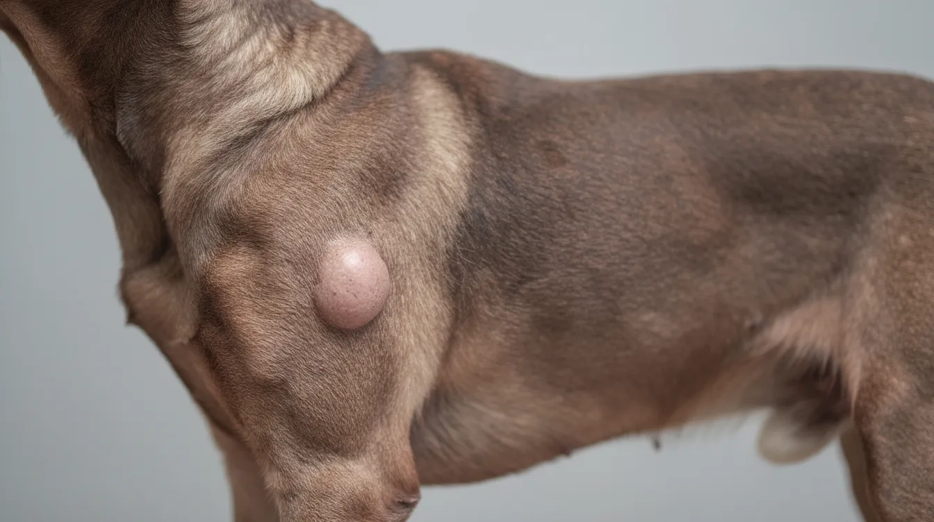
Lipomas (Fatty Tumors)
In some cases, bumps on dogs skin can become symptomatic, indicating a need for veterinary assessment.
Lipomas are benign fatty tumors common in older, overweight dogs. These growths form from fat cells beneath the dog’s skin and are among the most frequently diagnosed skin lumps in veterinary medicine. Lipomas are common in older, overweight dogs and grow slowly-typically over months or years.
Lipomas appear as soft, round or oval masses that are freely movable beneath the skin. Size varies dramatically-from small marble-sized lumps to large masses that can interfere with a dog’s movement. Certain dog breeds show higher predisposition, including Labrador Retrievers, golden retriever lines, Doberman Pinschers, Miniature Schnauzers, and Weimaraners. While lipomas themselves are benign, a rare malignant variant called liposarcoma exists, which is why veterinarians commonly recommend a fine needle aspirate to confirm the diagnosis.
Veterinarians will often discuss bumps on dogs skin with pet owners to understand the dog’s health history.
Skin Tags
It is essential to report any changes you notice in bumps on dogs skin to your veterinarian immediately.
Skin tags are small, narrow, stalk-like growths of fibrovascular tissue that hang from the skin surface. They are typically the same color as surrounding skin and develop in areas where skin rubs against skin or where friction from a collar or harness occurs. Common locations include the chest, armpits, and neck folds.
Skin tags are generally painless and rarely problematic unless they become irritated, snagged, or begin to bleed. They are most common in older dogs and do not require treatment unless they cause discomfort. Complete removal through minor surgery is straightforward if needed.
Concerning Bumps Requiring Veterinary Attention
The following growths require prompt professional evaluation because they may be malignant. Even if a bump looks similar to a benign growth, any rapid changes in appearance warrant a veterinary visit for fine needle aspiration or biopsy.
Mast Cell Tumors
Mast cell tumors are the most common malignant skin tumors in dogs, accounting for approximately 7–21% of all skin tumors. These growths originate from mast cells-immune cells involved in allergic reactions and inflammation. What makes them particularly dangerous is their extraordinary visual variability, earning them the nickname “the great imitators.”
Identifying bumps on dogs skin early can lead to better treatment outcomes and less stress for both the pet and the owner.
Mast cell tumors can appear as raised or flat bumps, pink or pigmented, firm or soft. A hallmark feature is intermittent swelling-the tumor may swell and then shrink as it releases histamine, sometimes causing intense itching or redness in the surrounding area. Typical locations include the chest, abdomen, and limbs. Breed predispositions are significant: Boxers, Boston Terriers, Bulldogs, Pugs, and Labrador Retrievers face elevated risk. The median age of diagnosis is around 10 years, though mast cell tumors can develop at any age. Diagnosis requires grading via biopsy, and treatment usually involves surgical removal with wide margins, potentially followed by radiation therapy or chemotherapy for high-grade or metastatic disease.
Melanomas
Malignant melanomas originate in melanocytes-the pigment-producing skin cells. Malignant melanomas commonly develop on the lips and mouth, under nail beds, and on pigmented skin areas. They typically present as dark brown or black raised bumps, though some are amelanotic (non-pigmented), which complicates visual detection.
Key visual warning signs include irregular borders, uneven color distribution, rapid growth, and ulceration. Melanomas affecting the mouth or nail beds are particularly aggressive with high metastatic potential. Surgical removal is the primary treatment, with adjunct therapies having limited effectiveness for advanced disease. Any new dark bump-especially on the lips, mouth, or near nail beds-should prompt immediate veterinary evaluation.
Hemangiosarcomas
Angiosarcomas (hemangiosarcomas) are highly malignant blood vessel tumors in dogs. On the skin surface, they appear as red, blood-filled bumps resembling blood blisters or bruised-looking patches. They are most common in areas with less fur and greater sun exposure, including the abdomen and inner thighs.
These tumors are extremely aggressive and can rupture, causing internal or external bleeding. Light-colored, short-haired dog breeds with significant sun exposure face higher risk. Any red, blood-filled skin lump that appears suddenly should be evaluated immediately by a veterinarian.
Step-by-Step Visual Examination and Fine Needle Aspiration Guide
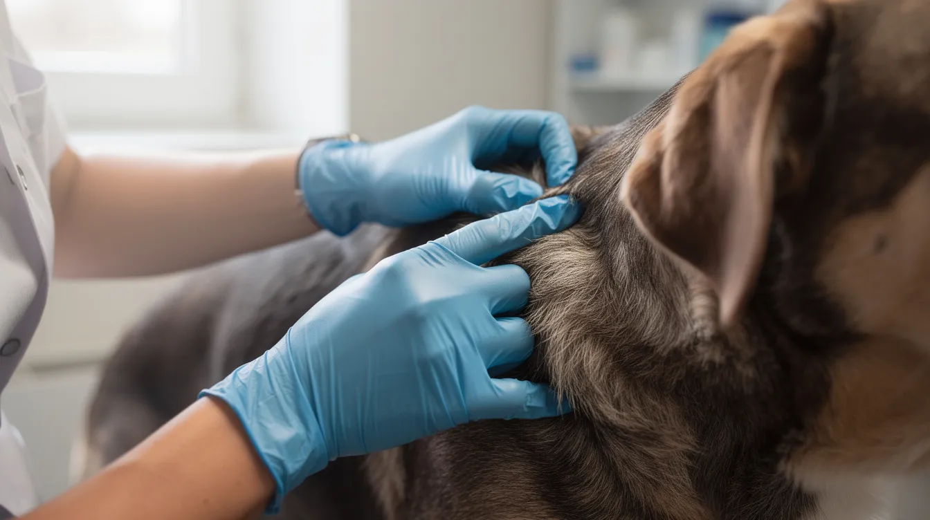
Performing a thorough at-home examination helps you gather valuable information before your veterinary appointment. Here is how to safely assess bumps on your dog’s body:
- Proper positioning and lighting: Place your dog in a comfortable position under good lighting-natural daylight is ideal. Part the dog’s coat to fully expose the bump and surrounding skin. Check for any missing fur, scaly skin, or flaky skin around the growth.
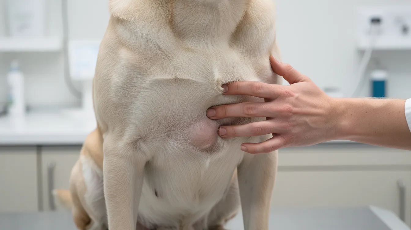
- Safe palpation technique: Using clean hands, gently feel the bump. Note whether it is soft or firm, freely movable or fixed to underlying tissues, tender or painless. Check if your dog flinches, pulls away, or shows discomfort. A soft, movable lump is more consistent with benign growths, while firm, fixed masses that do not move under the skin may indicate a problem.
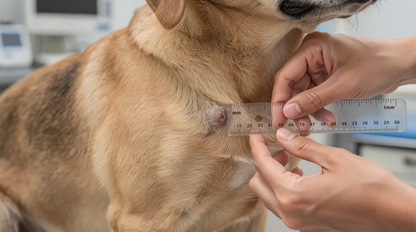
- Documentation methods: Photograph the bump from multiple angles-straight-on, from the side, and close-up. Place a ruler or coin next to the bump for scale. Record the date, exact location on the dog’s body, approximate size in centimeters, color, and texture. This documentation is invaluable for tracking changes over time.
- Reference comparison: Compare your photos and observations against the visual descriptions in this guide. Note similarities and differences, but remember that various benign and malignant lumps can appear similar-visual comparison alone cannot replace professional diagnosis.
Red Flag Visual Indicators
The following comparison helps distinguish normal bump characteristics from concerning features:
Feature | Likely Benign | Potentially Malignant |
|---|---|---|
Growth Rate | Slow (months to years) | Rapid (weeks), sudden appearance |
Texture | Soft, freely movable | Firm, fixed to deeper tissue |
Surface | Smooth, may be hair-covered | Ulcerated, crusted, bleeding |
Color | Skin-tone, uniform pink | Irregular pigmentation, dark or mixed colors |
Pain | Usually painless | Painful, warm, causes licking |
Borders | Well-defined, round | Irregular, poorly defined edges |
Size Changes | Stable or very slow growth | Doubles in size within weeks |
Rapid growth of a lump on a dog is a cause for concern and should trigger an immediate veterinary visit. Watch especially for bumps that ulcerate, bleed spontaneously, or show color changes over days to weeks.
When to Seek Veterinary Care
Understanding urgency levels helps pet parents respond appropriately when they discover skin bumps on their dogs.
Immediate Veterinary Attention Required
Seek same-day veterinary care when a bump is rapidly growing, actively bleeding, or causing your dog visible pain. Lumps in sensitive areas-including the mouth, nail beds, around the eyes, or near joints-need immediate veterinary attention regardless of appearance. Other urgent indicators include: swelling that fluctuates in size (suggesting mast cell involvement), discharge of pus (indicating possible abscesses, which are painful swellings filled with pus), interference with eating, breathing, or movement, and any bump showing signs of a secondary skin infection such as redness, warmth, or foul odor.
Routine Veterinary Consultation Recommended
Monitoring the appearance of bumps on dogs skin should be part of your regular pet care routine.
Schedule a veterinary appointment within 1–2 weeks for any new bump that has appeared recently but remains stable. This includes growths on older dogs or breeds predisposed to cancerous tumors, any change in an existing bump’s size, color, or texture, and situations where multiple small lumps have appeared. Even stable-looking growths in dogs over 7–8 years old warrant professional evaluation, as the incidence of both benign and malignant skin conditions increases with age. Perianal adenomas are common in older, unneutered male dogs and should be evaluated to rule out malignancy.
Home Monitoring Acceptable
Consulting resources about bumps on dogs skin can be beneficial for understanding potential health issues.
Home monitoring is reasonable for bumps with confirmed benign features-such as a classic soft, movable lipoma previously examined by a vet-that have been stable for 6 or more months, cause no pain, and do not interfere with function. Take photographs every 2–4 weeks and measure the growth to document any changes. If any change occurs in size, color, texture, or your dog’s behavior toward the bump, upgrade to a veterinary consultation.
Conclusion and Next Steps
This visual guide equips you with the knowledge to recognize common skin bumps on dogs and understand their general characteristics. However, visual identification is only the first step-it cannot replace veterinary diagnosis through fine needle aspiration, biopsy, and histopathology. Many cancerous tumors mimic benign growths, and even experienced veterinarians rely on laboratory analysis for definitive answers.
If you have identified a concerning bump on your dog, take these immediate steps:
- Document the bump with photographs, measurements, and notes on texture and mobility
- Schedule a veterinary appointment-same-day for urgent red flags, within 1–2 weeks for stable new growths
- Avoid squeezing, popping, or applying home remedies to any undiagnosed lump
- Monitor for additional skin problems such as hair loss, hot spots, scaly skin, or signs of skin disease elsewhere on the dog’s body
Treatment options range from watchful waiting for benign growths to surgical removal, radiation therapy, chemotherapy, or combination approaches for malignant tumors. The underlying cause and grade of the growth determine the treatment path. Diagnostic costs for a fine needle aspirate typically run $150–$500, while treatment for malignant growths including surgery and adjunct therapy may range from $1,000 to $3,500 or more depending on tumor type, location, and the dog’s size.
Early detection consistently improves outcomes. Make regular skin checks part of your grooming routine-parting the dog’s coat and running your hands over the entire dog’s body-so you catch new growths early and give your veterinarian the best chance at successful treatment.
Frequently Asked Questions
Discussing bumps on dogs skin with fellow pet owners can also provide valuable insights into what to expect.
Can I diagnose my dog’s bump from pictures alone?
No. Pictures help with initial recognition and can guide the urgency of your response, but they cannot substitute for a veterinary examination. Various benign and malignant lumps can appear similar, and cellular-level analysis through fine needle aspiration or biopsy is required for definitive diagnosis. Even trained veterinarians cannot reliably distinguish all tumor types by appearance alone.
How quickly do cancerous bumps typically grow in dogs?
Growth rates vary by tumor type. Some malignant growths double in size within weeks, while others progress more slowly. Rapid growth of a lump on a dog is a cause for concern and warrants prompt veterinary evaluation. Mast cell tumors add complexity because they can swell and shrink due to histamine release, making growth patterns appear inconsistent.
What should I do if I find a new bump on my dog?
Observe and document the bump immediately: take photos with a size reference, note the location on the dog’s body, and assess texture and mobility through gentle palpation. Schedule a veterinary appointment, especially if the bump shows any red flag features. Do not attempt to lance, squeeze, or remove the growth at home, as this can cause infection or spread cancerous cells.
Are certain dog breeds more prone to developing skin bumps?
Yes. Boxers, Boston Terriers, Bulldogs, and Pugs face higher risk for mast cell tumors. Labrador Retrievers, Miniature Schnauzers, Doberman Pinschers, and Weimaraners are more prone to lipomas. Light-colored, short-haired dog breeds with greater sun exposure have increased risk for squamous cell carcinomas. Breeds with dense or oily coats are more susceptible to sebaceous cysts and other skin conditions.
When is bump removal surgery necessary for dogs?
Surgical removal is recommended when a bump causes discomfort, interferes with movement or eating, is ulcerated or bleeding, is suspected or confirmed malignant, or is located in a sensitive or critical area. For cancerous tumors, complete removal with wide surgical margins is typically the primary treatment, and for some malignant masses surgeons remove enough skin around the area to improve the chance of complete excision. Operations near the anal sphincter can carry a risk of fecal incontinence, so surgical planning depends heavily on tumor location. Benign growths may also be surgically removed if they grow large enough to affect quality of life.
How can I tell if a bump is painful for my dog?
Consider joining community forums discussing bumps on dogs skin to share experiences and advice with other dog owners.
Watch for behavioral cues: flinching when the area is touched, persistent licking or chewing at the bump, swelling or warmth around the growth, reluctance to be handled, or changes in activity level. Some dogs may not show obvious pain signs even with uncomfortable growths, so a veterinary examination including palpation can better assess whether pain is present.
What’s the difference between a cyst and a tumor on dogs?
Cysts are encapsulated structures formed by blocked sebaceous glands or hair follicles, filled with fluid or semi-solid material like keratin. They tend to have smooth surfaces and well-defined borders. Tumors are proliferative cell masses that may be benign or malignant-malignant tumors can invade surrounding normal tissue and metastasize to organs. Cysts may discharge material if ruptured, while malignant tumors may ulcerate and bleed. Both require veterinary evaluation to confirm their nature.
Should I be worried about multiple small bumps on my senior dog?
Multiple small lumps on older dogs are common and frequently benign-lipomas, skin tags, and sebaceous gland tumors are commonly found in older dogs, but not every bump is a cyst, tumor, or other age-related growth. However, the presence of multiple benign growths does not rule out malignancy in any individual bump, and prolonged sun exposure can sometimes contribute to multiple tumors over time. Sebaceous gland tumors are common in older dogs and may bleed if irritated. Some bumps are infection-related, such as superficial pyoderma, and bacterial infections can cause folliculitis in dogs. Some senior dogs also develop greasy or flaky lesions tied to secondary seborrhea, while primary seborrhea is hereditary and less common. Itchy, inflamed patches may instead reflect food allergies, acute moist dermatitis, or acral lick dermatitis rather than a true lump. Each new or changing growth should be individually assessed by a veterinarian, and regular skin checks are especially important for senior dogs.
Additional Resources
- Merck Veterinary Manual – Comprehensive resource covering tumors of the skin in dogs, including diagnostic approaches, breed predispositions, and treatment protocols for both benign and malignant growths.
- Cornell University College of Veterinary Medicine – Offers veterinary dermatology educational materials, diagnostic flowcharts, and image libraries emphasizing the importance of cytology and histopathology in distinguishing skin conditions that affects dogs.
- American College of Veterinary Dermatology – Professional association providing specialist referrals for complex skin disease cases, including other skin diseases such as atopic dermatitis, demodectic mange, fungal infection, and yeast infections called malassezia dermatitis, plus differentials like mange called scabies caused by microscopic mites, external parasites, contact dermatitis, primary seborrhea, secondary seborrhea, ear infections, and food allergies; workups may also involve medicated shampoos, medicated baths, or oral drugs depending on the diagnosis.
Reviewed by Dr. Roger Hart, DVM, for medical accuracy and current veterinary best practices.
Leave a Reply